Head-to-Toe Nursing Assessment: Complete Examination, Documentation and Abnormal Findings Guide
Learn adult head-to-toe nursing assessment with system checklists, abnormal findings, documentation examples, clinical cases and practice rationales.
1. Quick Answer and Essential Facts
A head-to-toe nursing assessment is a systematic examination that combines the patient's account, observation, physical findings, vital signs and relevant records. Its purpose is to recognize current needs, establish a useful baseline, detect change and decide what requires nursing action or escalation. It is not a race through body parts. A patient with an immediate threat needs an emergency response before a routine checklist is completed.
This guide focuses on a general adult assessment in hospital and supervised learning settings. Pediatric, maternity, emergency, critical-care and specialist examinations require additional competencies and age- or condition-specific standards. A student should perform techniques within the placement's scope, supervision and consent requirements. The order below is a teaching framework; it does not replace an approved assessment tool or local deterioration pathway.
The most useful assessment answers five questions: What is usual for this person? What is happening now? What has changed? What could cause harm if missed? What needs to happen next? A normal-looking number cannot answer those questions alone. A person's report of new breathlessness, weakness or chest pressure can make an otherwise ordinary observation urgent.
| Assessment element | What it contributes | A common mistake |
|---|---|---|
| Patient's account | Symptoms, priorities, function and baseline | Replacing the patient's words with an assumption |
| Observation | Appearance, interaction, breathing and movement | Looking only at the monitor |
| Physical examination | Findings obtained with appropriate technique | Recording a finding that was not assessed |
| Measurements | Values that can be compared over time | Omitting position, device, oxygen support or units |
| Clinical reasoning | Priorities, further assessment and response | Completing the checklist despite deterioration |
| Reassessment | Evidence of response or continuing risk | Treating documentation as the end of care |
Remember: “Head to toe” describes organization. Patient condition determines priority.
The Open RN adult assessment checklist provides a useful entry-level reference for routine examination, with focused assessment and escalation when findings are unexpected. This guide uses original explanations, examples and practice cases rather than reproducing that checklist.
Go deeper: Build the foundation with fundamentals of nursing complete guide, nursing care plans assessment diagnosis interventions, vital signs for nurses.
2. How to Use This Guide for Clinical Learning
Begin with the general survey and emergency-priority sections, then learn one body-system examination at a time. For every technique, connect the action to a purpose. Listening to lung fields is more useful when you can explain what asymmetry, diminished airflow or a new adventitious sound would change about your next action. Memorizing the movement of the stethoscope without understanding the question produces a performance rather than an assessment.
Use three passes when studying. First, understand the expected finding and the limitations of the technique. Second, rehearse the sequence with a consenting classmate or simulator under the required supervision. Third, practise interpreting a changed finding and communicating it. That third pass is where many learners discover that recognizing an abnormality and responding to it are different skills.
Keep a short learning log. Record the technique, what you found difficult, the feedback received and the specific improvement to practise. Do not include patient identifiers. “I need to compare corresponding posterior lung fields without listening through clothing” is more actionable than “I am bad at assessment.” The log should guide deliberate practice, not become another lengthy document that is never reviewed.
For assignments, distinguish an educational example from a real patient record. The sample documentation in this guide is fictional. It illustrates clarity and reasoning, not a template to copy into a chart. A real record must reflect what the nurse actually assessed and did.
Go deeper: Connect assessment findings to the nursing process and ADPIE, then practise the NCJMM clinical-judgment steps.
3. Comprehensive, Focused, Ongoing and Emergency Assessment
A comprehensive assessment builds a broad picture. It may be appropriate at admission, a major transition or a planned full examination. It includes history and function as well as physical findings. An ongoing assessment compares current status with that baseline and with the previous assessment. The amount of detail needed depends on the setting, patient acuity and what has changed.
A focused assessment investigates a particular concern. New dizziness might require attention to symptoms, blood pressure, pulse, medication timing, glucose when indicated, hydration and neurologic findings. It does not mean ignoring the rest of the patient. The nurse first checks for immediate danger and then gathers the information most likely to clarify the problem.
An emergency assessment is organized around threats that cannot wait. If someone becomes unresponsive or is not breathing normally, the nurse activates the appropriate emergency pathway and performs trained emergency actions. It would be unsafe to continue a routine pupil or bowel-sound examination while postponing that response. The full examination can be completed once urgent needs are addressed.
| Type | Main question | Example |
|---|---|---|
| Comprehensive | What is the overall baseline and care need? | Admission history with a general physical assessment |
| Ongoing | What is different from the established baseline? | Shift assessment after a medication change |
| Focused | What explains this specific concern or treatment response? | Reassessment of breathing after prescribed therapy |
| Emergency | What immediate threat must be addressed now? | Sudden collapse or severe respiratory distress |
These categories overlap. A routine assessment can become a focused examination and then an emergency response. The ability to change direction is a sign of good judgment, not failure to follow the sequence.
4. Preparation Before Entering the Room
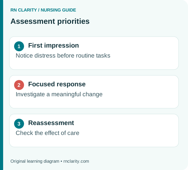
Review the reason for admission, relevant history, allergies, current precautions, recent observations and important treatment changes. Look for the patient's usual cognitive and functional status, oxygen requirements, mobility instructions and communication needs. A brief review should orient the examination without replacing direct assessment. Information in the record can be outdated, copied forward or incomplete.
Check that necessary equipment is available and working. A routine assessment may require a stethoscope, appropriate blood-pressure cuff, thermometer, timing device, penlight and protective equipment. Additional equipment depends on the concern and local practice. Clean reusable equipment and perform hand hygiene at the appropriate moments. Follow transmission-based precautions before entering the designated patient space.
Plan practical needs that affect participation. The patient may need glasses to read a scale, a hearing aid to follow instructions, an interpreter to describe symptoms accurately or toileting before a longer examination. A tired or breathless person may need shorter stages. A patient in pain may require comfort measures or prescribed analgesia and reassessment before particular movements are tolerable.
Avoid deciding in advance that the examination will be normal because the previous nurse documented stability. Use previous findings as a comparison, not a conclusion. Equally, do not assume that a chronic abnormality is a new emergency without clarifying the baseline. Preparation should make your questions more precise and your examination safer.
5. Identification, Consent, Privacy and Communication
Introduce yourself and your role, verify the patient using the organization's approved identifiers, and explain what you intend to do. Room number or bed position is not a reliable identifier. Ask how the person prefers to be addressed and whether they are comfortable with visitors remaining. Someone who agrees to a general examination may still want privacy for particular questions or body areas.
Explain before touching, exposing or repositioning. Consent is an ongoing conversation. A patient can ask you to stop or decline a part of the examination. Explore the concern respectfully, explain the purpose and possible consequences of an incomplete assessment, and involve the appropriate clinician when needed. Do not label a refusal as noncooperation without understanding pain, fear, trauma history, cultural needs or communication barriers.
Use a qualified interpreter when needed according to policy. Speak directly to the patient and allow enough time for responses. A family member may provide valuable baseline information, but their account should be identified as collateral information rather than silently substituted for the patient's own report. Document who supplied important information when that distinction affects interpretation.
A practical opening is: “I will ask about how you are feeling, check your vital signs and examine the areas relevant to your care. Please tell me if anything is painful or if you want a break.” This gives the patient a role in the assessment and makes unexpected discomfort easier to report.
6. The First Look: General Survey and Immediate Safety
Observe before concentrating on equipment. Can the patient engage in conversation? Is breathing comfortable? Is their position chosen for comfort or because lying flat causes distress? Do they appear newly drowsy, agitated, pale, sweaty or markedly weak? These observations do not establish a diagnosis, but they help determine how urgently you must investigate.
Compare what you see with what was expected. A patient who was talking normally an hour ago and now answers only after repeated stimulation has changed, even if the monitor displays a reassuring oxygen saturation. A patient who is chronically unable to speak needs a communication approach suited to that baseline; silence alone is not evidence of reduced consciousness.
Check the immediate environment. Note oxygen delivery, visible lines, drains, alarms, mobility supports and hazards. Confirm that tubing is not creating a trip risk or being pulled by the patient's position. Environmental assessment is part of patient assessment because equipment and surroundings can contribute to harm or explain a symptom.
A safe opening sequence
- Observe responsiveness, breathing, overall appearance and obvious distress.
- Identify any immediate threat and activate the appropriate response.
- Confirm identity, explain the assessment and support communication.
- Establish the patient's main concern and usual baseline.
- Proceed systematically while remaining ready to change priorities.
Do not wait for a perfectly complete set of observations before obtaining help for a visibly deteriorating patient. Gather additional information while the response is being mobilized when this can be done safely.
7. Build a Symptom History That Guides the Examination
Ask an open question first: “What feels different today?” Then clarify onset, location, quality, severity, timing, associated symptoms and what makes the concern better or worse. A symptom framework such as PQRST can help organize questions, but the letters should not force an awkward interview or delay urgent care.
Separate onset from recognition. A patient may have noticed weakness at breakfast but last been known at their normal baseline the previous evening. That distinction can matter in time-sensitive neurologic evaluation. Similarly, “short of breath since yesterday” needs clarification about whether symptoms were gradual, suddenly worse, present at rest or related to a specific activity.
Ask about function, not only symptom intensity. “Can you walk to the bathroom as you usually do?” may reveal a meaningful decline that a broad “Are you okay?” misses. Explore sleep, appetite, elimination, falls, medication access and the patient's ability to manage usual activities when relevant. These details connect the physical examination to actual care needs.
Record uncertainty honestly. If the patient cannot recall when a symptom began, document that the onset is uncertain and identify any reliable collateral information. Do not convert an estimate into an exact time. The clinical team needs an accurate account of what is known, what is reported and what remains to be clarified.
8. Vital Signs: Technique, Context and Trends
Vital signs are interpreted observations, not isolated numbers. Record the measurement method and context that could influence the result. Blood pressure depends on cuff size and position; temperature depends partly on device and site; oxygen saturation must be understood alongside oxygen support, signal quality and the patient's condition. Compare a current value with previous values obtained under reasonably similar conditions.
For a resting adult, common orientation ranges include a pulse of approximately 60–100 beats per minute and respiratory rate of approximately 12–20 breaths per minute. These are reference points, not universal definitions of safety. Fitness, medications, age, illness and individual baseline affect interpretation. A newly slow pulse with dizziness or a rising respiratory rate with fatigue may be important even before a threshold is crossed.
The MedlinePlus vital-sign overview provides general adult reference information. The FDA's pulse-oximeter guidance emphasizes device limitations. Pulse oximetry estimates oxygen saturation; it does not directly measure ventilation or exclude carbon-dioxide retention.
| Observation | Context worth recording | Interpretation question |
|---|---|---|
| Blood pressure | Position, arm, cuff and relevant symptoms | Is this accurate, expected and adequate for this patient? |
| Pulse | Rate, rhythm, method and symptoms | Is the rhythm regular, and has it changed? |
| Respirations | Rate, depth, effort and pattern | Is breathing becoming less effective or more difficult? |
| Temperature | Value, device/site and trend | Does the pattern fit the clinical concern? |
| Oxygen saturation | Device, oxygen support and signal | Does the reading agree with the patient's appearance? |
Go deeper: Use the vital-signs guide for measurement technique, orthostatic assessment and trend interpretation.
9. Pain and Discomfort Assessment
Pain assessment starts with the patient's report when they can communicate reliably. Ask about location, quality, onset, pattern, severity, associated symptoms and impact on function. A numeric score can track change, but it does not replace the history or examination. A patient with a modest pain score and new chest pressure may need more urgent evaluation than someone with a higher score from an unchanged, already assessed condition.
Use an appropriate validated observational tool when the patient cannot provide a reliable self-report, following local practice and the population for which the tool is intended. Absence of speech does not mean absence of pain. Conversely, agitation, grimacing or tachycardia can have causes other than pain. Interpret behavioral cues with context rather than treating a tool score as a diagnosis.
Assess safety before and after analgesic interventions. Sedation, respiratory pattern, blood pressure and other observations may be relevant depending on the medication and patient. Reassessment should establish whether the intervention improved comfort or function and whether adverse effects appeared. “Medication administered” describes an action; it does not describe the outcome.
Ask what a meaningful improvement would look like. A patient may value being able to cough, sleep or reposition more than reaching a score of zero. Document the agreed functional goal where appropriate. New, unexplained, rapidly worsening or disproportionate pain requires assessment and escalation; it should not automatically be attributed to the known diagnosis.
10. Mental Status, Orientation and Neurologic Baseline
Observe alertness, attention, speech, behavior and ability to follow appropriate instructions. Orientation questions help describe cognition, but a patient can answer their name and location correctly while still showing an important change in attention or behavior. Use the assessment tool required in your setting and compare findings with the person's usual function.
Establish the baseline with the patient, previous documentation and appropriate collateral history. New confusion in an older adult should not be dismissed as normal aging or automatically labeled dementia. Delirium has multiple possible contributors, including acute illness, medications, pain, hypoxia, metabolic disturbance and unfamiliar surroundings. The nursing priority is to recognize the change, assess immediate safety and seek evaluation of reversible causes.
When a standardized scale such as the Glasgow Coma Scale is used, document its component findings and factors that limit assessment according to policy. Intubation, language barriers, hearing loss, sedation and pre-existing deficits affect how a response should be interpreted. Do not give a patient a seemingly precise score by guessing an untestable component.
For a newly reduced level of consciousness, prioritize airway, breathing, circulation and urgent help. Check relevant causes within the local emergency pathway, including glucose when indicated. A detailed memory examination can wait. The key handoff information is the change from baseline, its timing, associated findings and actions already taken.
Practical check: “Normally converses independently; now needs repeated verbal stimulation to answer” communicates a clinically useful change. “Sleepy” alone leaves important questions unanswered.
11. Pupils, Facial Movement, Strength and Sensation
Assess pupil size and reaction using appropriate lighting and a penlight when indicated. Compare both eyes and note relevant baseline conditions, surgery or medications. Unequal pupils may be longstanding or new; the distinction matters. New asymmetry accompanied by headache, reduced consciousness or focal neurologic findings requires urgent evaluation rather than a routine note for the next shift.
Observe facial symmetry, speech and spontaneous movement. Compare corresponding limbs for strength and movement using the technique taught and required in your setting. Ask about numbness or tingling, and assess sensation when relevant. A painful joint, immobilization device, prior stroke or language barrier may limit performance. State the limitation instead of treating inability to complete a maneuver as proof of a new neurologic lesion.
Sudden facial weakness, arm weakness or speech difficulty should trigger the applicable stroke response. Establish the last-known-well time when possible and communicate it promptly. Do not delay escalation to finish a complete cranial-nerve examination. The American Stroke Association warning-sign resource supports prompt recognition and emergency response.
Recheck significant findings and seek help without creating avoidable delay. Comparing two observations can distinguish a technique problem from a true change, but repeated testing is not a substitute for escalation when the patient is clearly deteriorating. Document what was observed rather than a diagnosis that has not been established.
12. Head, Eyes, Ears, Nose, Mouth and Neck
A general examination includes the areas relevant to the patient's symptoms, function and care. Ask about changes in vision or hearing, dizziness, headache, oral discomfort and swallowing problems. Check whether glasses, dentures or hearing devices are needed and available. An apparent communication or cognitive problem may partly reflect missing sensory aids.
Inspect accessible areas with consent and suitable lighting. Note visible lesions, swelling, drainage, mucosal moisture, oral hygiene and device-related pressure. Avoid interpreting a single finding beyond its reliability. Dry mouth can be associated with medications, oxygen therapy, mouth breathing or reduced intake; it is not a stand-alone diagnosis of dehydration.
Ask about swallowing and observe relevant cues. Coughing, choking, a wet-sounding voice or difficulty managing secretions warrants assessment through the local swallowing pathway. Do not use an informal drink test when the patient has a condition requiring a validated screen or specialist assessment. Oral intake and medication routes may need review before administration.
Neck examination may include inspection and additional techniques appropriate to competence and setting. Do not palpate both carotid arteries simultaneously. A general nursing assessment does not require every advanced maneuver taught in a specialist physical-examination course. Choose techniques that answer a relevant question and that you are trained and authorized to perform.
Communication findings can change the examination
- Difficulty hearing: check hearing support and use an accessible communication approach.
- Difficulty seeing: provide glasses or another suitable aid before interpreting task performance.
- Difficulty speaking: establish whether this is baseline or new, and assess urgently if new.
- Difficulty swallowing: follow the swallowing-safety pathway before oral intake when indicated.
13. Respiratory Inspection Before Auscultation
Watch the patient breathe before asking them to take repeated deep breaths. Note rate, rhythm, depth, effort, posture, ability to speak and symmetry of chest movement. A person who can say only a few words at a time may be working hard to breathe even if the oxygen-saturation reading is not dramatically low. Fatigue and reduced effort after a period of severe distress can be ominous rather than reassuring.
Ask about cough, sputum, breathlessness, chest discomfort, usual oxygen use, respiratory diagnoses and recent treatment. Establish whether the patient is on room air or supplemental oxygen and record the device and prescribed settings as appropriate. An oxygen saturation without that context is an incomplete comparison.
Look for equipment issues that can explain a change without assuming equipment is the whole problem. A disconnected oxygen tube needs attention, but the patient still needs assessment. A poor pulse-oximeter signal may be technical, while new breathlessness can be real at the same time. Check both the person and the system supporting them.
Do not diagnose the cause of respiratory distress from appearance alone. Wheeze, crackles, reduced breath sounds and tachypnea can occur in different conditions. The general assessment identifies the pattern and urgency; further evaluation establishes the cause. Immediate nursing actions follow the patient's condition, authorized protocols and orders.
14. Lung Auscultation and Interpretation
Use the stethoscope directly on skin where appropriate, maintain privacy and compare corresponding areas from side to side. Help the patient position safely. If sitting forward is not tolerated, adapt the examination and document the limitation. Do not force a breathless or unstable person through an elaborate sequence solely to complete a student checklist.
Listen for airflow and unexpected sounds, noting where they occur and whether they change after coughing or repositioning when such reassessment is appropriate. Describe findings accurately: a new unilateral reduction in breath sounds is different from generally quiet sounds in a patient with a known chronic condition. The distribution and change from baseline matter.
| Finding | What to clarify | Safe interpretation |
|---|---|---|
| Wheeze | Location, severity, work of breathing and response to therapy | Suggests narrowed airflow but does not identify a single cause |
| Crackles | Distribution, newness, cough, oxygen need and fluid context | May accompany several pulmonary or cardiac problems |
| Reduced sounds | Symmetry, technique, baseline and associated symptoms | New asymmetry or distress requires prompt evaluation |
| Stridor | Onset, airway symptoms and overall stability | Potential upper-airway threat requiring urgent response |
| Apparently normal sounds | Symptoms, respiratory effort and trend | Do not exclude important disease on auscultation alone |
If you cannot confidently identify a sound, describe what you hear and seek supervised confirmation. Inventing a label makes the record less useful. Clinical learning improves when uncertainty is made explicit and then resolved with feedback.
Go deeper: Link respiratory observations to ABG interpretation and the vital-signs guide.
15. Cardiovascular Symptoms, Pulse and Heart Sounds
Ask about chest discomfort, palpitations, dizziness, syncope, breathlessness, exercise tolerance and swelling. Clarify whether symptoms are new, changing or usual. A patient who describes “indigestion” may still have a time-sensitive problem; use the full history and clinical context rather than relying on the patient's label.
Assess the pulse using the method required for the situation. Note rate, rhythm, strength and relevant symptoms. An irregular pulse may require an apical assessment, rhythm monitoring or further evaluation according to policy. A normal rate does not prove adequate circulation, and an abnormal rate does not establish its cause.
When auscultating the heart, identify the expected sounds and any finding that requires clarification. Use the locations and technique you have been taught, avoiding unsupported claims about a murmur or extra sound. If a new sound is accompanied by breathlessness, chest symptoms or hemodynamic change, communicate the whole pattern rather than only the acoustic finding.
Compare the examination with medications and recent procedures. A new slow pulse after a rate-lowering medication, or new chest symptoms after a procedure, provides context that can change urgency. The nurse should not independently invent a medication-hold threshold; follow the order, assess symptoms and clarify an unsafe situation promptly.
The value of this part of the examination lies in connecting electrical activity, mechanical circulation and the patient's experience. A monitor rhythm is one source of information, not a replacement for palpating a pulse and assessing perfusion when indicated.
16. Peripheral Perfusion, Edema and Vascular Concerns
Compare corresponding extremities for temperature, color, pulses, sensation, movement and swelling where appropriate. Interpret these findings together. A cool limb, new pain, altered sensation and reduced pulse is a different situation from longstanding bilateral ankle edema. Sudden or progressive changes in perfusion or neurovascular status need prompt escalation through the relevant pathway.
Capillary refill is one observation of peripheral circulation. Temperature, lighting, technique and patient factors can affect it. Do not use a single refill time as proof that circulation is normal or abnormal. If you use a grading scale for pulses or edema, follow the organization's definitions and describe the relevant location and comparison. A grade without a site or trend is hard to interpret.
For edema, note whether it is unilateral or bilateral, dependent or generalized, new or chronic, and associated with pain, skin change or respiratory symptoms. Edema can have different causes and can coexist with reduced effective circulating volume. Do not assume that swollen ankles automatically mean the patient needs more diuretic or that all prescribed fluid must be stopped.
Unilateral calf swelling or pain warrants assessment for possible venous disease and other causes. Do not massage a limb when thrombosis is suspected, and do not rely on a provocative maneuver such as Homan's sign to diagnose or exclude DVT. The appropriate diagnostic pathway depends on clinical evaluation and ordered testing. New breathlessness or chest symptoms in this context increases urgency.
Perfusion is a pattern
- Central clues: mental status, blood pressure, pulse and overall appearance.
- Peripheral clues: temperature, color, pulses, refill and limb symptoms.
- Organ clues: urine output, respiratory status and other findings relevant to the patient.
- Trend: what changed, how quickly, and after which intervention?
17. Abdominal Assessment: History and Examination Order
Ask about abdominal pain, nausea, vomiting, appetite, swallowing, distension and bowel pattern. Clarify the last bowel movement and what is normal for the patient. Ask about blood, black stool or a change in stool appearance when relevant. Medication use, recent surgery, reduced mobility and diet changes may help explain symptoms, but they should not be used to dismiss new or severe findings.
For a routine abdominal examination, the commonly taught sequence places inspection and auscultation before percussion and palpation, because manipulating the abdomen may affect bowel sounds. Follow the examination taught for your role and setting. Use gentle technique and avoid repeating painful maneuvers unnecessarily. The presence of severe pain, rigidity, hemodynamic instability or another acute concern should change the plan and prompt appropriate escalation.
Bowel sounds are only one part of the examination. Their presence does not prove that oral intake is safe or exclude obstruction. Their apparent absence should be established using the local assessment method rather than a brief listen followed by a definitive label. Decisions about feeding, imaging or treatment require the wider clinical picture and the appropriate clinician's assessment.
Describe the patient's location of pain and your observed findings separately. “Reports pain in the right lower abdomen; guards during gentle examination” is more defensible than diagnosing a specific condition from one bedside sign. Do not perform deep palpation over a suspected pulsatile abdominal mass; escalate the concern and follow the appropriate pathway.
18. Nutrition, Hydration and Swallowing Function
Assessment includes whether the patient can obtain, tolerate and safely consume the planned nutrition. Ask about appetite, recent intake, nausea, difficulty chewing, swallowing concerns and unintended weight change. Observe the practical barriers: a meal placed out of reach, dentures that do not fit, breathlessness while eating or difficulty opening containers can all reduce intake despite an adequate diet order.
Interpret hydration with several findings. Intake, output, weight trend, symptoms, blood pressure, pulse, mucous membranes, medications and relevant laboratory results may contribute. Skin turgor or dry mouth alone is insufficient, particularly in older adults. Edema does not guarantee adequate circulating volume. Fluid assessment is a clinical interpretation, not a single physical sign.
For patients with enteral access, assess the visible site, securement, device markings when relevant and the prescribed feeding arrangement. Verify placement and safe use through the approved device-specific process. Do not use air insufflation and abdominal listening as proof of feeding-tube position. A displaced or uncertain tube requires clarification before use according to policy.
When swallowing safety is uncertain, follow the established screening and referral pathway. The route for fluids, food and medications may need review. Explain the reason for any temporary restriction respectfully and provide appropriate mouth care and comfort measures. A patient who is thirsty should not be left without an explanation or a plan.
Go deeper: Review fluid and electrolyte assessment and safe medication administration when intake, fluid balance or route changes affect treatment.
19. Urinary Assessment and Urine-Output Interpretation
Ask about usual voiding, recent frequency, urgency, pain, difficulty starting, incomplete emptying and incontinence. Determine when the patient last voided and whether their pattern has changed. A report of “not passing urine” may represent low production, retention, an unrecorded void or a collection problem. Those possibilities lead to different assessments.
For a catheterized patient, inspect the system without unnecessary disconnection. Check for visible kinks, dependent loops, traction, securement problems or a drainage bag positioned inappropriately. Assess urine characteristics and the amount collected, then compare with the clinical picture. Cloudy or odorous urine alone does not establish a symptomatic infection or justify antibiotics.
Low urine output can accompany reduced perfusion, obstruction, kidney dysfunction and other problems. Assess the patient and the collection system, review trends and escalate according to the setting's thresholds and the overall condition. Do not treat a single low hourly amount by automatically increasing fluids without an order or protocol, particularly when congestion or kidney disease may be present.
If a bladder scan or other assessment is indicated, use it within competence and policy. Record the method, result and relevant symptoms. When reporting low output, include the duration, recent intake, blood pressure, pulse, bladder or catheter findings, and actions already taken. “Urine low” gives the receiving clinician too little information to judge urgency.
The CDC catheter-associated urinary tract infection recommendations support appropriate indications, maintenance of a closed system and prompt removal when a catheter is no longer needed.
20. Skin Assessment Across Skin Tones
Inspect the skin with consent, adequate lighting and attention to pressure areas, folds and sites under medical devices. Assess color in the context of the person's usual skin tone, and combine visual findings with temperature, tenderness, texture and tissue consistency. Early pressure-related damage may be harder to recognize by redness alone in darker skin tones.
Look at areas vulnerable to pressure and moisture, including heels, sacral regions and sites beneath oxygen tubing, masks, splints or other devices. Reposition safely with appropriate assistance. A complete skin assessment should not create a fall, line dislodgement or respiratory problem. If an area cannot be assessed at that time, document the reason and arrange a safe opportunity to complete it.
Distinguish pressure injury from other skin conditions, including moisture-associated damage, skin tears and vascular wounds. Use the current staging framework and seek specialist help when classification is uncertain. Not every pressure injury fits a simple stage-one-to-four description: unstageable injury, deep tissue pressure injury and mucosal pressure injury require appropriate terminology. Do not assign a stage by guessing what lies beneath obscuring tissue.
Describe location, appearance and relevant measurements using the organization's wound-assessment method. Record pain, drainage, surrounding skin and any device contribution. A photograph may supplement assessment only through approved consent, privacy and documentation processes; a personal phone is not an appropriate shortcut.
Refer to the National Pressure Injury Advisory Panel staging resources for the classification definitions. Prevention and management require an individualized plan rather than a stage label alone.
21. Mobility, Strength, Balance and Functional Safety
Ask what the patient normally does independently and what help they currently need. Assess the activities relevant to the care plan: turning, sitting, standing, transferring, walking or using an assistive device. A person who can move both legs in bed may still be unsafe to stand because of dizziness, weakness, pain or impaired balance.
Check current activity orders and precautions before testing movement. Recent surgery, fractures, weight-bearing restrictions, hemodynamic instability and other conditions may limit what is safe. Use the prescribed assistance level and appropriate equipment. Do not ask a patient to demonstrate walking alone merely to see whether they can manage.
Observe quality as well as completion. Does the patient become breathless, lightheaded or markedly fatigued? Do they need to pull on furniture, lose balance or forget instructions? Does pain change the way they transfer? These findings can influence fall prevention, toileting plans, rehabilitation referral and discharge readiness.
Reassess after changes that may affect mobility, including sedating medications, diuresis, an acute illness or prolonged bed rest. A previous “independent” status is not permanent. Communicate the current safe assistance level clearly so that other staff do not rely on an outdated assumption.
The CDC STEADI resources provide structured fall-risk assessment resources for older adults. Their use should complement clinical judgment and the tools required in the specific care setting.
22. Orthostatic Symptoms and Safe Position Changes
Ask about dizziness, blurred vision, weakness or near-fainting when sitting or standing. Review relevant medication changes, fluid losses and the patient's ability to stand safely. Orthostatic blood-pressure assessment is not an exercise to complete regardless of symptoms. A person who becomes unstable should be supported and the test stopped or modified according to clinical judgment and policy.
The CDC STEADI orthostatic assessment uses five minutes lying down, a supine blood pressure and pulse, then measurements after one and three minutes of standing. A fall in systolic pressure of at least 20 mm Hg or diastolic pressure of at least 10 mm Hg within three minutes is a commonly used definition of orthostatic hypotension. Symptoms and safety remain important even when a numerical threshold is not met.
Document positions and timing rather than listing three unexplained blood pressures. Include the patient's symptoms, assistance used and whether the procedure was completed. If standing was unsafe, state that rather than reporting an incomplete set as a negative result.
A symptom during transfer may require a broader assessment than orthostatic measurements alone. New chest discomfort, focal weakness, an irregular pulse or persistent altered consciousness can indicate a different or additional problem. Do not let a plausible explanation become a reason to ignore other cues.
23. Lines, Drains, Dressings and Medical Devices
Identify every device relevant to the patient's care and understand its purpose. Trace lines carefully and compare the observed arrangement with the order and documentation. Assess visible insertion sites, securement, tubing, drainage or infusion, and the patient's symptoms. A general assessment should reveal whether a device is functioning safely and whether its continued need is being reviewed.
For an IV site, look for pain, swelling, leakage, erythema, temperature change and other signs of complication. A pump running normally does not prove that the catheter is patent or that the medication is reaching the intended space. If infiltration or extravasation is suspected, follow the device- and medication-specific response; do not automatically flush a painful swollen site.
For drains and dressings, record the relevant amount, character and trend of output or visible drainage. Do not remove a dressing or manipulate a device merely because it appears on a checklist if an order, procedure-specific instruction or local policy limits that action. Examine safely and obtain clarification when the necessary assessment cannot be completed within the current instructions.
Device documentation should be specific enough for continuity of care. “Lines okay” does not identify which line was checked, where it is located or what was observed. Record the findings that matter for that device and setting, including concerns, actions and follow-up.
Go deeper: See IV therapy for nurses and infection prevention for device complications, aseptic practice and monitoring principles.
24. Psychosocial Assessment and Patient Priorities
Ask how the patient is coping and what concerns them most. Hospitalization can disrupt sleep, independence, family roles, finances and the sense of control. These concerns can affect participation, symptom reporting and discharge readiness. They belong in a nursing assessment when they influence care, rather than being treated as an optional conversation after physical tasks are finished.
Observe distress without assuming its cause. Anxiety may coexist with hypoxia, pain, medication effects or another acute problem. A person who appears withdrawn may be tired, depressed, overwhelmed, unable to hear or communicating in an unfamiliar language. Explore the finding and assess the relevant physical and psychosocial possibilities.
Use respectful, direct questions when a safety assessment is indicated, including questions about self-harm, abuse or an unsafe discharge environment. Follow the organization's validated process, privacy safeguards and escalation requirements. Do not promise secrecy when information must be shared to protect safety or meet applicable obligations.
Include strengths. Ask what usually helps, who the patient trusts, what they can manage independently and what support they want involved. A care plan built only around deficits misses opportunities for effective partnership. Record the patient's stated priorities in language that preserves their meaning and avoids judgmental labels.
For an assignment, explain how a psychosocial finding changes the plan. “Worried about affording medication; referred for discharge support and confirmed the follow-up plan” is more useful than a generic statement that emotional support was provided.
25. Adaptation for Older Adults and People With Disability
Do not equate older age with confusion, frailty or dependence. Establish individual baseline and preferences. Some older adults are highly active and independent; others have complex support needs. New loss of function can be an important sign of acute illness even when classic symptoms are less obvious.
Allow time for responses and accommodate hearing, vision, fatigue and mobility needs. Distinguish a limitation in the assessment method from a limitation in the patient. For example, inability to read small print is not evidence of poor understanding, and inability to hear an instruction is not evidence of impaired attention. Use accessible formats and verify understanding.
Ask people with disability how they usually perform activities and what adaptations work for them. A familiar transfer technique or communication system may be safer than an unfamiliar approach chosen without consultation. Follow current mobility and device instructions, and collaborate with the appropriate team members when specialist support is needed.
Document baseline deficits and new changes separately. “Chronic right-sided weakness unchanged from reported baseline” and “new reduction in left-hand grip” communicate different risks. Avoid using a broad label such as “abnormal neurologically” when a precise description is possible.
The goal is an accurate and equitable assessment. Adaptation is not lowering the standard; it is choosing a method that makes the findings interpretable for the person in front of you.
26. When to Stop the Routine Assessment
Stop or redirect the sequence when findings suggest immediate danger. Examples include severe breathing difficulty, new reduced responsiveness, signs of acute stroke, sudden severe chest symptoms, uncontrolled bleeding, a threatened airway or rapidly worsening circulation. Activate the local emergency or rapid-response process and perform trained, authorized actions.
The nurse does not need a final diagnosis before escalating a concerning change. A clear statement of the problem and evidence is enough to request urgent evaluation. “New confusion, respiratory rate 30, falling blood pressure and reduced urine output” is a meaningful report even before the cause is confirmed.
| Situation | Why the routine sequence changes | What to communicate |
|---|---|---|
| New difficult arousal | Airway, ventilation or other acute causes may threaten safety | Baseline, onset, respiratory findings, medications and response |
| Sudden focal weakness or speech change | Time-sensitive neurologic evaluation is needed | Last known well, observed deficits and associated findings |
| Severe dyspnea or stridor | Breathing or airway support may be needed immediately | Effort, rate, oxygen support, saturation and progression |
| Rapidly worsening perfusion | Shock or another urgent process may be developing | Blood pressure, pulse, mental status, skin and output trends |
| Unexpected major bleeding | Ongoing blood loss can cause rapid deterioration | Site, estimated/observed loss, symptoms and relevant therapies |
Do not spend excessive time obtaining repeated “better” measurements while urgent care is delayed. A quick technical check can be appropriate, but it should occur alongside a timely response to the patient.
From finding to action
- Recognize a significant new finding or trend.
- Assess immediate airway, breathing, circulation and safety needs.
- Activate the appropriate level of help.
- Provide the observations, timing and relevant background.
- Carry out authorized interventions and reassess the response.
- Document the event and ensure ongoing responsibility is clear.
27. Turn Observations Into a Nursing Problem
An assessment becomes useful when findings are organized into a supported problem or risk. Start by clustering related cues. Breathlessness, increasing oxygen need, crackles and reduced activity tolerance may form one clinically important pattern. New dizziness, a medication change and unsafe standing may form another. Keep the clusters open to revision as more information becomes available.
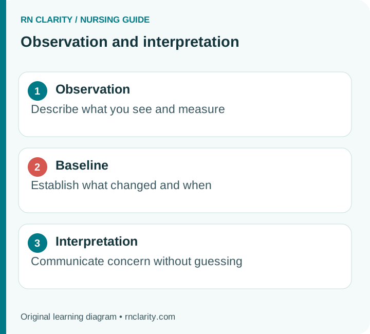
Distinguish observations from interpretations. “Respiratory rate 28 with accessory-muscle use” is an observation. “Breathing is ineffective” is an interpretation that should be supported by the wider assessment. A medical diagnosis may explain the situation, but the nursing plan still needs to identify the patient's current response and care needs.
Prioritize by immediate threat, instability, time sensitivity and patient-specific risk. Do not apply a mnemonic mechanically. A routine educational need may be important but should not take priority over a new breathing problem. A risk may deserve urgent prevention even before harm occurs, such as unsafe swallowing in a patient about to receive oral medication.
For care-plan assignments, use the terminology and diagnostic framework required by the program. Verify that each diagnosis is supported by the assessment and that each goal is measurable. Do not attach a long list of diagnoses to every patient simply because those labels appear in a textbook chapter.
Go deeper: The nursing diagnoses guide and care-plan guide explain how to connect cues, problems, goals, interventions and evaluation.
28. Documentation: Describe What You Actually Assessed
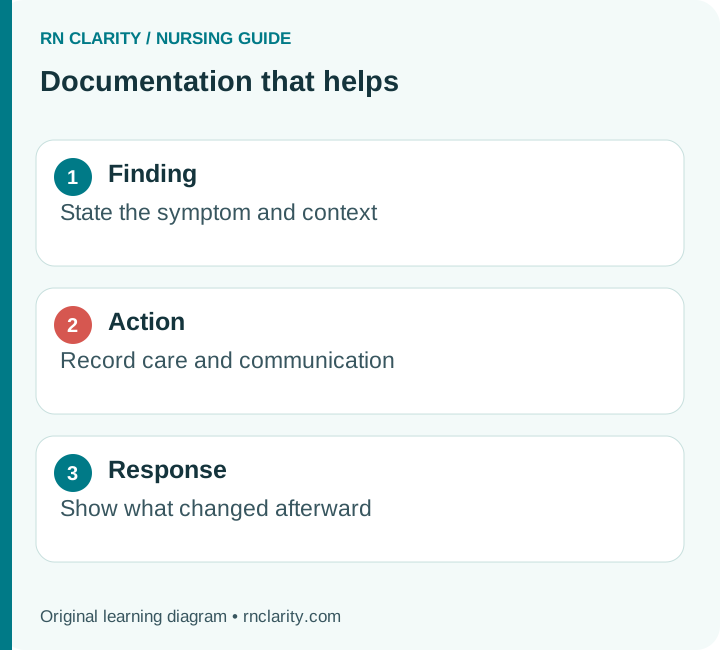
Chart promptly using the approved record and terminology. Include relevant objective findings, the patient's report, limitations, important changes, interventions, communication and response. Avoid blanket phrases that suggest a more complete examination than you performed. “Within normal limits” is only meaningful when the organization's defined assessment has actually been completed and the statement accurately applies.
Use the patient's words for a symptom when their wording is clinically important. Separate reported information from observed findings. “Patient reports no urine since breakfast” differs from “No urine measured in the collection device since 0800.” Both may be true, but they answer different questions.
Record limitations and follow-up plans. If the patient declines a skin examination or cannot tolerate turning because of respiratory distress, state the reason and what was done to address the incomplete assessment. Do not fill a template with normal findings to avoid blank fields. Missing data honestly identified is safer than invented reassurance.
Document significant communication with enough detail to show that responsibility and follow-up are clear. Note the concern, person contacted, relevant instructions and response according to policy. An unanswered page does not establish that the problem has been addressed; use the escalation process when necessary.
28.1 A fictional documentation comparison
Too vague: “Patient fine. Lungs okay. Up to bathroom.”
More useful: “Patient alert and conversing; denies new breathlessness at rest. Respiratory pattern unlabored on prescribed oxygen support. Assisted to bathroom using current mobility plan; reported lightheadedness on standing, returned safely to sitting. Blood pressure/pulse reassessed and findings escalated according to unit protocol.”
The second example still requires actual values and details in a real record. It demonstrates a relationship between the finding, action and response rather than providing a complete chart entry to copy.
29. SBAR and Handoff: Make the Priority Easy to Understand
SBAR organizes a conversation into situation, background, assessment and recommendation or request. It is a communication aid, not a script that must be completed before urgent help is activated. Start with the concern and urgency. A receiving clinician should not have to listen through a long history before learning that the patient has become difficult to arouse.
Give only the background needed to interpret the immediate problem. Include relevant diagnoses, procedures, medications, baseline and recent changes. State the assessment findings with times and trends where possible. Explain what you have already done and whether the patient improved, worsened or remained unchanged.
Make the request explicit. “Please review the patient now,” “I need clarification before this medication is administered,” and “Please confirm the monitoring and escalation plan” are clearer than ending with “Just letting you know.” Use read-back or closed-loop communication when required for critical information and orders.
A concise escalation report
- Situation: What is happening now, and how urgent is it?
- Background: What baseline, diagnosis or treatment change matters?
- Assessment: What did you observe, measure and reassess?
- Request: What review, clarification or immediate support is needed?
At handoff, include unresolved concerns and tasks that still require follow-up. A complete examination does not eliminate the need to communicate the one abnormality that matters most. Confirm that the next clinician understands pending results, reassessment times and the current assistance level.
30. Reassessment and Evaluation After an Intervention
Choose reassessment measures that correspond to the intervention and the problem. After a breathing intervention, evaluate respiratory effort, symptoms, oxygenation and other relevant observations. After assistance with orthostatic symptoms, assess the patient's stability and safe mobility plan. After analgesia, evaluate comfort, function and medication-specific safety findings.
Timing depends on the clinical situation, intervention, route and local protocol. There is no single reassessment interval suitable for every medication or patient. A deteriorating person needs immediate ongoing observation and response, while a stable patient receiving routine care may follow a scheduled plan. State when reassessment is due and who is responsible if care is transferred.
Do not assume that an intervention worked because it was completed. A nebulizer may have been administered while respiratory effort continues to worsen. A fluid order may have been delivered without an improvement in perfusion. A patient may report less pain while becoming excessively sedated. Evaluation requires both intended effects and possible adverse effects.
If the outcome is not achieved, reconsider the assessment and escalate as appropriate. Avoid repeating the same intervention automatically without examining why it failed. The nursing process is iterative: new findings may change the problem, goal or treatment plan. Document the response and the resulting next step so that the record shows a coherent episode of care.
31. A Repeatable Bedside Assessment Checklist
Use this checklist as a memory aid after learning the underlying techniques. It does not replace the required assessment form, population-specific tools or a clinician's examination. The order can change when symptoms, positioning, precautions or fatigue make another sequence safer. A person who cannot tolerate lying flat should not be forced flat simply to complete a checklist in its printed order.
| Checkpoint | What to establish | What must carry forward |
|---|---|---|
| Opening | Identity, consent, communication needs, precautions and immediate distress | Any barrier requiring an interpreter, equipment or assistance |
| General survey | Appearance, interaction, position, breathing and stated concern | The patient's priority and any urgent finding |
| Neurologic | Current mental status, relevant baseline, movement and focused findings | New deficits, timing and actions already taken |
| Cardiopulmonary | Symptoms, vital-sign trends, respiratory effort, sounds and perfusion | Oxygen device, ordered target and concerning changes |
| Gastrointestinal | Intake, symptoms, abdomen and bowel pattern as indicated | Aspiration concerns, intolerance or new abdominal findings |
| Genitourinary | Voiding symptoms, output context and device need | Retention concerns, reduced output or catheter issues |
| Skin and movement | Exposed skin, pressure areas, wounds, assistance and function | Protection measures and current mobility plan |
| Devices | Indication, site, securement, settings and pathway as appropriate | Any mismatch, malfunction or unresolved concern |
| Closing | Comfort, position, access to help and explanation of next steps | Reassessment, escalation and handoff responsibilities |
Before leaving, ask whether the patient has an additional concern that has not been addressed. A person may wait until the end to disclose dizziness during toileting, trouble paying for a medication or fear about discharge. Those concerns can materially change a care plan even when the physical findings appear stable.
The checklist is complete only when abnormal findings have an appropriate response. Recording that a dressing is saturated without communicating the change is an incomplete clinical response. Likewise, documenting an unsafe transfer without arranging assistance leaves the risk unresolved. Connect each relevant finding with a next step, a responsible person and an evaluation plan.
32. Case Study: A New Neurologic Change During Routine Care
Fictional learning scenario. A hospitalized adult conversed clearly during handoff. During the morning assessment, the nurse notices new difficulty naming familiar objects and reduced movement of one arm. The patient says the symptoms began recently but cannot provide an exact time. A visitor reports that the patient spoke normally during a conversation earlier that morning.
The priority is to recognize a possible acute neurologic emergency and activate the facility's stroke or emergency response process. Do not finish the abdominal and skin examination first. Obtain or communicate the last-known-well information, current findings and relevant observations without delaying help. Assess airway, breathing, circulation and immediate safety while the response team is mobilized. Check point-of-care glucose when indicated by the emergency protocol because glucose abnormalities can contribute to neurologic symptoms; a result does not remove the need to evaluate a persistent focal deficit.
The nurse should avoid assuming a specific stroke subtype from bedside findings. Brain imaging and the treating team's evaluation guide time-sensitive treatment. Oral food, fluids and medications require attention to swallowing safety and the emergency plan. A previously normal swallow does not guarantee that swallowing remains safe after a new deficit.
Reasoning to practice: What changed, compared with what baseline, at approximately what time? Which observations will help the team act immediately? Which routine activities should pause? The strongest handoff leads with the new deficit and its timing, then adds relevant context. A long recital of unrelated normal findings can obscure the emergency.
Evaluation: Document the recognition time, response activation, available last-known-well information, relevant findings and transfer of care. Do not write that a stroke was confirmed before the diagnostic team establishes it. The nursing contribution is rapid recognition, accurate communication and safe support during evaluation.
33. Case Study: Sedation After an Analgesic
Fictional learning scenario. A postoperative patient reports improved pain after an opioid but now drifts to sleep during conversation. Breathing appears shallow, and the patient is harder to awaken than at the previous assessment. The oxygen saturation is within the ordered target while supplemental oxygen is running.
The saturation value must not override the change in consciousness and ventilation. Assess responsiveness, respiratory rate and quality, airway patency and other immediate findings. Activate the appropriate urgent response and follow the facility's opioid-related deterioration protocol. Further sedating medication requires review. Support ventilation and administer reversal treatment only within authorized protocols and orders, with the required monitoring and assistance.
This case illustrates why pain reassessment includes safety. A lower pain score is not a successful outcome when the patient has developed clinically concerning sedation. Supplemental oxygen can also make oxygen saturation an incomplete indicator of ventilation. The nurse should communicate the medication, dose, route and timing from the actual administration record, together with changes in alertness and breathing.
Reasoning to practice: Which cue is the most important change? What information identifies a possible medication contribution without prematurely excluding other causes? How will the patient's response be reassessed after emergency treatment? The presence of an opioid does not prove that every episode of deterioration is an opioid overdose, so ongoing assessment remains necessary.
Evaluation: Continue observation according to the emergency plan, including recurrence of sedation or respiratory compromise. A brief improvement does not establish sustained recovery. Document the findings and response, then contribute to a revised analgesia and monitoring plan that supports both comfort and safety.
34. Case Study: Dizziness During the First Walk
Fictional learning scenario. An adult who has spent much of the day in bed becomes light-headed on standing. The patient grips the bedside table and says, “I need to sit down.” The nurse was preparing to assess walking distance.
The immediate response is to assist the person to a safe position and obtain help as needed. Do not insist on completing a standing measurement or walking test while the patient is unstable. Assess symptoms and relevant vital signs, then communicate concerning or persistent findings. Potential contributors include medication effects, volume changes and other illnesses, but the bedside observation alone does not establish the cause.
Once safe, review baseline mobility, recent intake and losses, medication timing, current restrictions and the need for a more focused evaluation. Orthostatic measurements may be appropriate when ordered or permitted by the assessment protocol. Record the position and timing of each measurement so the results can be interpreted. “Blood pressure low” does not communicate whether a value was obtained supine, sitting or standing.
Reasoning to practice: The task has changed from measuring walking performance to preventing a fall and evaluating symptoms. The plan may need a different assistance level, a gradual position change or further clinical review. Do not label the person noncompliant because the walk was stopped for symptoms.
Evaluation: Reassess symptoms before another transfer, update the assistance plan and tell the next caregiver what happened. The patient should know how to request help and why independent walking is temporarily unsafe. Successful assessment includes this practical change in care, not merely a completed mobility score.
35. Case Study: A Device Conceals a Skin Problem
Fictional learning scenario. During an assessment, a nurse finds a tender area beneath an oxygen-device strap. The surrounding skin looks different from the patient's adjacent skin, and the patient reports that the device has felt tight for several hours. Oxygen support is still clinically required.
Assess the affected area using the appropriate skin and pressure-injury process, including the person's symptoms, temperature, tissue consistency and visible findings. Skin changes may be harder to recognize by color alone in darker skin tones. Do not remove essential oxygen support without maintaining the ordered therapy; obtain help to relieve device pressure safely and select an appropriate alternative or adjustment.
Determine whether the finding needs specialist assessment and follow the reporting process. Pressure injury, friction injury, moisture-associated damage and other conditions are not interchangeable labels. Document the location and objective findings rather than guessing a stage from an incomplete view. A wound covered by slough or eschar may not permit determination of full depth, and mucosal pressure injuries are not staged using the skin staging system.
Reasoning to practice: What part of the assessment was only possible after examining beneath or around a device? How can the source of pressure be addressed while preserving treatment? What information should be included in the next device and skin check?
Evaluation: Reassess comfort, device function and the affected tissue after the intervention. Ensure that the revised plan is communicated so another caregiver does not tighten the strap back to the previous position. This is an example of assessment preventing an ongoing exposure rather than simply recording existing damage.
36. Case Study: Confusion Is Not Automatically Dementia
Fictional learning scenario. An older adult with a documented history of dementia is unusually sleepy, has difficulty sustaining attention and cannot participate in activities that were manageable yesterday. A family member says, “This is very different from normal.”
Treat the acute change as a new clinical problem. Compare the current presentation with a reliable baseline and assess immediate physiologic concerns. Review relevant observations, medication exposure, pain, hydration and other possible contributors while arranging appropriate clinical evaluation. Use the facility's validated delirium assessment process when trained and indicated. A chronic cognitive diagnosis does not explain away a sudden change.
Adapt communication by using short statements, allowing response time and ensuring that hearing and visual aids are available when appropriate. Avoid assuming that a person who cannot provide a detailed history has no pain or symptoms. Family and caregivers can contribute useful baseline information, but the nurse should still address the patient directly and preserve dignity.
Reasoning to practice: Which findings suggest a change in attention or level of arousal? What was the person able to do previously? Which reversible or urgent problems need evaluation? The assessment should distinguish observed behavior from labels such as “difficult” or “uncooperative.”
Evaluation: Track changes over time and communicate the assessment conditions. A brief period of clearer conversation does not necessarily mean that the underlying problem has resolved. Record the patient's function and attention, the support provided and the clinical follow-up plan.
37. Turning Cases into Clinical Judgment Practice
Cases become more useful when learners must explain the connection between a cue and an action. Start with a short description and identify the change from baseline. Then name what additional information would help without delaying an urgent response. Decide which problem deserves attention first and explain why. Finally, specify what improvement or deterioration would look like on reassessment.
- Notice: Identify a meaningful new finding.
- Interpret: Compare the finding with symptoms, context and baseline.
- Prioritize: Decide whether routine care can continue safely.
- Respond: Take authorized action and communicate.
- Evaluate: Check the patient's response and revise the plan.
For a small-group exercise, assign one learner to describe objective findings, another to explain the priority and another to give a concise handoff. Ask the group what evidence would change its interpretation. This reduces the tendency to attach a diagnosis to the first familiar cue. A learner should be able to say, “I am concerned about this possibility, but further evaluation is needed,” while still acting promptly on a dangerous presentation.
Connect this exercise with the clinical judgment guide. In a clinical placement, follow the institution's emergency pathways and the supervising clinician's direction. Classroom reasoning supports practice but does not replace competency assessment or local procedures.
38. Practice Questions with Rationales
A patient develops new difficulty breathing halfway through the examination. What should the nurse do first?
Pause the routine sequence and assess the immediate breathing concern while obtaining the level of assistance required by the patient's condition. Completing the remaining systems before responding delays evaluation of a potentially urgent change. The priority is determined by the patient's presentation, not by the next item on a form. Document and communicate the relevant findings and response after immediate safety needs are addressed.
An automated blood pressure is very different from earlier readings. Can it be dismissed as equipment error?
No. Assess the patient and verify the measurement using correct technique and functioning equipment. A surprising number may reflect a technical problem, a real change or both. Symptoms and other signs guide urgency. Repeating a measurement should not delay emergency assistance when the patient appears unstable. Record the confirmed findings and relevant measurement conditions so the team can interpret the trend.
Why should a nurse ask about baseline function?
Baseline function makes a current finding interpretable. A mobility limitation present for years differs from a new inability to transfer. The same principle applies to speech, cognition, sensation and self-care. Baseline information does not make current limitations irrelevant; it helps distinguish chronic support needs from acute changes requiring further evaluation. State the source of the baseline information when documenting it.
Is “lungs clear” sufficient documentation for a patient with new breathlessness?
No. Breath sounds are only one part of respiratory assessment. The record should reflect relevant symptoms, respiratory rate and effort, oxygenation context, findings and actions. Clear auscultation does not rule out all serious causes of breathlessness. A useful note explains what the nurse observed, how the patient was supported and what evaluation or reassessment followed.
A patient declines part of the examination. How should this be handled?
Explain the purpose, explore the reason for declining and offer reasonable adaptations while respecting the patient's rights and applicable consent requirements. Pain, privacy concerns, fatigue, prior trauma and communication barriers may be addressable. Document what was offered, what was declined and any resulting limitation or follow-up. Do not chart a normal finding for an area that was not assessed.
Why should urine output be interpreted with timing and context?
An amount without a collection interval cannot describe a meaningful rate or trend. Consider intake, losses, body size when relevant, kidney history, symptoms and the accuracy of measurement. A catheter drainage problem may affect the recorded amount but should not automatically be assumed to explain reduced output. Persistent or concerning changes require assessment and appropriate communication.
A patient reports severe pain but appears calm. Which finding should be believed?
The patient's report is central when the patient can self-report. Outward behavior varies and does not reliably invalidate a report of pain. Assess location, characteristics, associated symptoms, functional effect and safety concerns, then follow the care plan. A calm appearance can be documented as an observation without using it to dismiss the experience or withhold appropriate evaluation.
Does a completed screening score replace clinical judgment?
No. Screening tools organize selected information and can trigger a standardized response, but they do not capture every relevant circumstance. A low score should not override a new alarming symptom. Use the tool correctly, interpret it with the rest of the assessment and follow required escalation criteria. Record the actual observations that prompted concern rather than relying solely on the score.
39. Frequently Asked Questions
What is a head-to-toe nursing assessment?
It is a systematic collection and interpretation of information about the patient's current condition, usually including a general survey, history, vital signs and examination of major body systems. The nurse compares findings with baseline, identifies concerns, communicates important changes and evaluates the response to care. It is more than a checklist and must adapt to the patient's condition and the clinical setting.
How long should a head-to-toe assessment take?
There is no universal time that defines a safe or complete assessment. A stable patient familiar to the nurse may need a different examination from a new admission with multiple concerns. Communication needs, mobility limitations and abnormal findings also affect duration. Efficiency comes from preparation and an organized sequence, while urgent findings may require the routine examination to stop.
What is the difference between a head-to-toe and a focused assessment?
A head-to-toe assessment provides a broad review of the patient's condition. A focused assessment examines a particular symptom, system or treatment response in greater depth. They complement each other. A routine examination may identify a concern that requires a focused follow-up, and a new urgent symptom may require a focused assessment before any broader examination can safely continue.
Which findings should be reported immediately?
Examples include new severe breathing difficulty, an acute neurologic deficit, loss of responsiveness, signs of major bleeding or other evidence of rapid deterioration. Use the facility's emergency criteria and clinical judgment. The urgency depends on the full presentation and change from baseline. Do not wait for every vital sign or a completed chart before activating help for an apparent emergency.
Should every patient have the same examination?
No. The core structure supports consistency, but the examination must reflect age, symptoms, diagnosis, treatment, functional ability, consent and setting. A routine adult inpatient checklist cannot replace a pediatric, obstetric, psychiatric or intensive-care assessment. Perform techniques within training and scope, and use population-specific tools when required. Explain adaptations and any limitations in the record.
Why is abdominal assessment usually taught in a different order?
Inspection and auscultation are generally performed before percussion and palpation because manipulation can influence bowel sounds and the patient's response. The exact depth of examination depends on training and clinical circumstances. New severe abdominal symptoms require appropriate escalation and further evaluation; a routine sequence or the presence of bowel sounds does not exclude a serious abdominal condition.
Can a nursing student use this guide during a skills checkoff?
It can support preparation and reasoning, but the program's rubric and instructor's directions determine the required performance. Practice technique under supervision and learn why each observation matters. Do not use a memorized script to ignore distress, consent or safety. A good checkoff demonstrates organized assessment, accurate communication and appropriate recognition of findings that need follow-up.
What should be documented when an assessment is incomplete?
Document the portions completed, the reason for the limitation, relevant findings and the plan to obtain missing information when appropriate. Examples include patient preference, severe fatigue, an urgent change or a positioning restriction. Avoid copying a normal template into an unassessed area. The record should make the limitation visible to the next clinician and identify any safety implications.
40. Using This Guide for Assignments and Further Study
For an assignment, separate descriptive nursing observations from diagnostic conclusions and evidence-based recommendations. A fictional case can demonstrate reasoning but cannot establish that an intervention improves outcomes. If your paper makes a treatment claim, consult the relevant current guideline or primary research and describe the population and limitations. Cite the original source when that is the evidence you are discussing.
Check the publication date and scope of every reference. A recently updated web page may summarize an older guideline, while an older foundational skills chapter may remain useful for basic technique. Neither date alone establishes quality. Ask whether the source addresses adults or children, routine care or emergencies, and the clinical environment in your assignment. Do not substitute an inpatient checklist for a specialty protocol.
When building a care plan, write from the patient's actual assessment rather than selecting a familiar label first. Identify supported problems, measurable goals and interventions linked to their rationale. Include an evaluation method that can show whether the patient's condition or function changed. Where your course requires a standardized nursing-diagnosis taxonomy, consult the licensed current edition and your institution's terminology requirements.
Practice next: Apply these ideas with the clinical judgment learning pathway and the practice-question study guide.
41. References and Source Notes
The following resources support the assessment methods and safety topics discussed here. Original cases, checklists and learning diagrams in this guide were created for education; they are not reports of actual patients or validated clinical decision tools.
- Open RN: Nursing Skills, second edition, Appendix C—Head-to-Toe Assessment Checklist. A foundational adult nursing-skills resource; adapt its checklist to the patient's setting and current local policies.
- MedlinePlus: Vital signs. General adult vital-sign context; reference ranges are not universal treatment targets.
- FDA: Pulse Oximeter Basics. Limitations of pulse oximetry and the importance of symptoms and clinical context.
- American Stroke Association: Stroke Symptoms. Sudden neurologic warning signs and the need for an emergency response.
- CDC STEADI: Measuring Orthostatic Blood Pressure. Standardized measurement technique and interpretation for the stated assessment context.
- CDC STEADI: Clinical Resources. Resources for assessment and prevention of falls in older adults.
- CDC: Summary of Recommendations for Prevention of Catheter-Associated Urinary Tract Infections. Appropriate catheter use and maintenance principles; the page summarizes the underlying guideline rather than representing a newly issued 2026 guideline.
- National Pressure Injury Advisory Panel: Pressure Injury Stages. Terminology for pressure-injury classification; use appropriate training and specialist consultation when findings are uncertain.
This guide is for nursing education and exam preparation. Clinical care must follow the patient's condition, current orders, professional scope and local protocols. RN Clarity's original educational content is not official NCLEX examination material and does not replace supervised clinical training.