NCLEX-RN® Eye and Ear Disorders: Glaucoma, Cataracts, Retinal Detachment, Hearing Loss, and Ménière Disease
Review high-yield eye and ear disorders, assessment findings, treatments, and nursing safety priorities.
postNumber: 88 title: "NCLEX-RN® Eye and Ear Disorders: Glaucoma, Cataracts, Retinal Detachment, Hearing Loss, and Ménière Disease" h1: "NCLEX-RN® Eye and Ear Disorders: Glaucoma, Cataracts, Retinal Detachment, Hearing Loss, and Ménière Disease" slug: "nclex-rn-eye-ear-disorders-glaucoma-retinal-detachment" canonicalUrl: "https://rnclarity.com/blog/nclex-rn-eye-ear-disorders-glaucoma-retinal-detachment" primaryKeyword: "NCLEX® eye and ear disorders" secondaryKeywords:
- "glaucoma NCLEX® nursing"
- "retinal detachment emergency signs"
- "cataract surgery nursing care"
- "hearing loss communication nursing"
- "Meniere disease NCLEX®" metaTitle: "NCLEX® Eye and Ear Disorders: Glaucoma and Hearing" metaDescription: "Learn glaucoma, cataracts, retinal detachment, eye emergencies, hearing loss, ear medications, Ménière disease, and nursing safety for NCLEX®." excerpt: "A clear sensory-disorders guide focused on emergency recognition, medication technique, postoperative safety, communication, and fall prevention." suggestedPublishDate: "2026-09-06" status: "draft_nurse_review_required" schemaTypes: ["BlogPosting", "FAQPage", "BreadcrumbList"] diagramCount: 5 clinicalReview: "Required before publication"
NCLEX-RN® Eye and Ear Disorders: Glaucoma, Cataracts, Retinal Detachment, Hearing Loss, and Ménière Disease
Quick answer: Sudden flashes, a shower of floaters, or a curtain-like shadow can signal retinal detachment and requires urgent eye evaluation. Sudden severe eye pain with halos, nausea, and a red eye can signal acute angle-closure glaucoma. Sudden hearing loss is also urgent. Protect the sensory organ, avoid pressure or delay, and communicate in a way the patient can use.
Editorial status: This is a complete educational draft. A qualified U.S. registered nurse or nurse educator should clinically review it before publication. Drug doses, facility procedures, and emergency algorithms must be checked against the current source and local policy.
Suggested reading time: 18–22 minutes
Designed for: NCLEX-RN® candidates, including internationally educated nurses and repeat test-takers.
Table of contents
- Recognize vision emergencies
- Understand open-angle and angle-closure glaucoma
- Care for cataracts and cataract surgery
- Recognize retinal detachment and macular problems
- Administer eye medications safely
- Communicate with hearing loss
- Understand Ménière disease, otitis, and vestibular disorders
- Administer ear medications and protect safety
- A simple NCLEX® clinical-judgment workflow
- Original practice scenarios with rationales
- Common NCLEX® traps
- What to memorize and what to understand
- A seven-day review plan
- Frequently asked questions
- Final rapid-review checklist
Why this topic matters for the NCLEX-RN®
Eye and ear questions test urgency and patient teaching. A gradual painless cataract is not managed like sudden painless vision loss. Chronic open-angle glaucoma is often silent and requires lifelong pressure-lowering treatment; acute angle closure is an emergency. Ménière disease creates vertigo and fall risk, while sudden sensorineural hearing loss needs rapid evaluation.
The NCLEX-RN® also tests how the nurse communicates with sensory impairment. Speaking loudly does not automatically help hearing loss, and shouting can distort words. Face the patient, reduce background noise, use clear speech, verify understanding, and make the environment safe.
Recognize vision emergencies
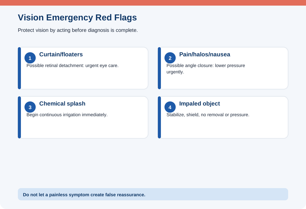
Treat sudden vision loss, eye trauma, chemical exposure, severe eye pain, new flashes and floaters, a curtain or shadow, and a fixed or irregular pupil after injury as emergencies. For chemical exposure, begin immediate irrigation with water or saline and continue while emergency evaluation is arranged; do not delay to identify the chemical. Remove contact lenses if they come out easily during irrigation.
For penetrating injury or an impaled object, do not remove the object or apply pressure. Stabilize it, shield both eyes to reduce sympathetic movement when appropriate, keep the patient from eating or drinking in case surgery is needed, and obtain emergency ophthalmic care. For blunt trauma, assess for vision change, pain, hyphema, globe rupture, and orbital injury.
A patient reporting painless transient vision loss can have retinal or vascular ischemia. Even if vision returns, urgent assessment is needed. Eye emergencies are time-sensitive because retinal and optic-nerve tissue may be permanently damaged.
| Finding | Possible emergency | First nursing priority |
|---|---|---|
| Flashes, floaters, curtain | Retinal tear/detachment | Urgent ophthalmic evaluation |
| Severe pain, halos, nausea, red eye | Acute angle closure | Immediate pressure-lowering treatment |
| Chemical splash | Corneal/ocular burn | Irrigate immediately |
| Impaled object | Open-globe injury | Do not remove; shield and stabilize |
| Sudden hearing loss | Sensorineural emergency | Urgent same-day evaluation |
Understand open-angle and angle-closure glaucoma
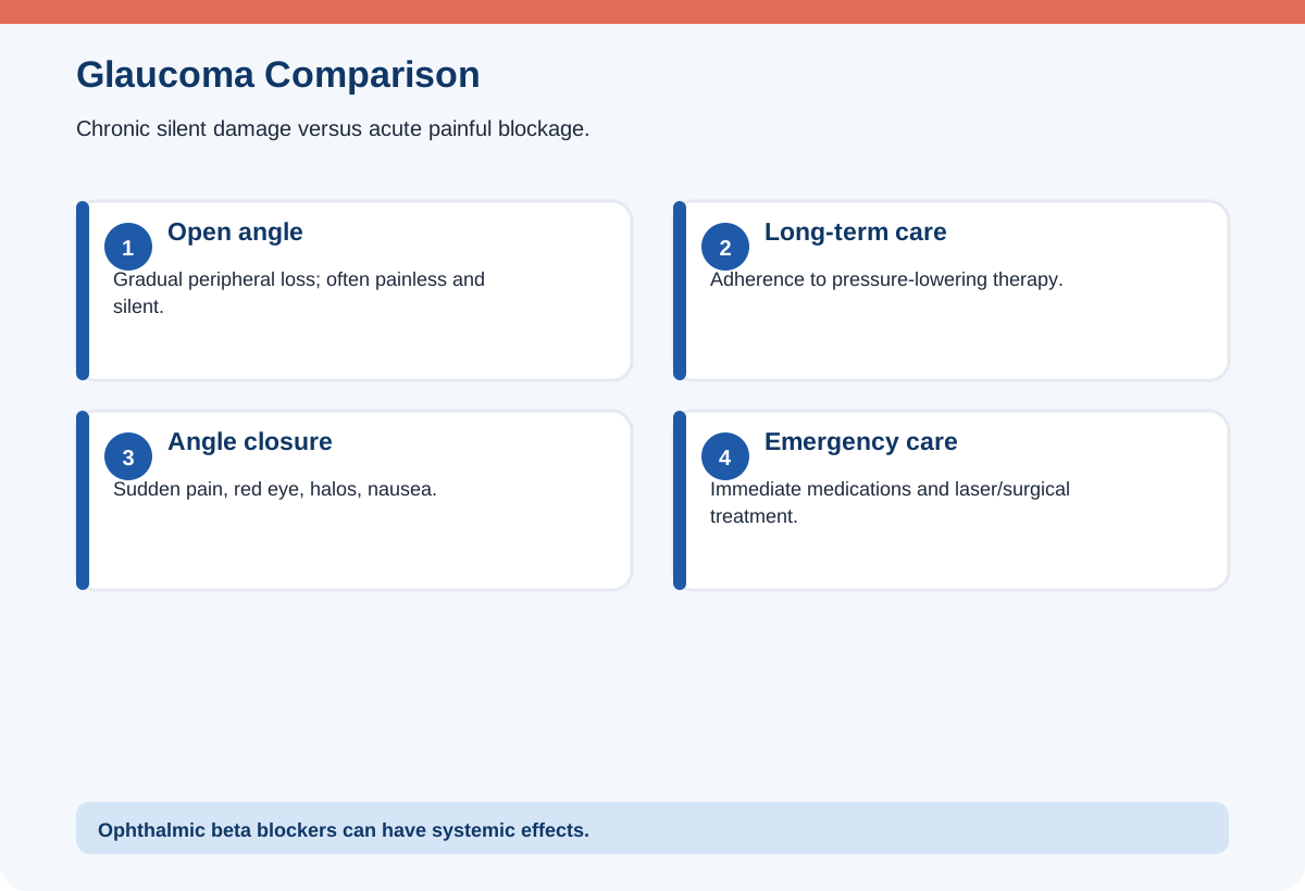
Glaucoma damages the optic nerve, often through elevated intraocular pressure or pressure susceptibility. Primary open-angle glaucoma usually develops slowly and painlessly, causing gradual peripheral-field loss. Because early symptoms may be absent, screening and adherence to lifelong eye-drop therapy are essential.
Acute angle-closure glaucoma causes a sudden blockage of aqueous drainage. Findings include severe eye pain, headache, blurred vision, halos, red eye, a mid-dilated poorly reactive pupil, nausea, and vomiting. It is an emergency requiring rapid medication and often laser or surgical treatment. Do not darken the room and wait; pupil dilation can worsen angle closure in susceptible eyes.
Common pressure-lowering drops include prostaglandin analogs, beta blockers, alpha agonists, carbonic anhydrase inhibitors, and cholinergic agents. Know systemic effects: ophthalmic beta blockers can worsen bradycardia, heart block, or bronchospasm. Use punctal occlusion to reduce systemic absorption.
Care for cataracts and cataract surgery
A cataract is clouding of the lens that causes gradual painless blurred vision, glare, reduced color clarity, poor night vision, and frequent prescription changes. Surgery removes the cloudy lens and places an artificial lens when symptoms interfere with function. Medication cannot dissolve a mature cataract.
After surgery, protect the eye, use drops exactly as prescribed, avoid rubbing, and follow restrictions on bending, heavy lifting, straining, or water exposure. Prevent constipation and forceful coughing when possible because sudden pressure may stress the surgical site. Wear the shield as directed, especially during sleep.
Report severe pain, sudden decreased vision, increasing redness, purulent drainage, flashes, floaters, or nausea. Mild scratchiness and some blurred vision may occur initially, but severe pain is not expected. Ensure fall precautions because depth perception and visual adjustment may temporarily change.
Recognize retinal detachment and macular problems
Retinal detachment may cause sudden flashes of light, new floaters, a dark curtain or shadow, and painless loss of part of the visual field. It requires urgent repair to preserve vision. Keep the patient safe, reduce activity as directed, and avoid food or drink when emergency surgery is possible. After repair with a gas bubble, strict head positioning may be ordered to keep the bubble against the retinal break.
A patient with an intraocular gas bubble must avoid air travel and high altitude until the bubble is absorbed because reduced atmospheric pressure can expand the gas and dangerously raise intraocular pressure. Nitrous oxide anesthesia is also contraindicated while an intraocular gas bubble is present.
Age-related macular degeneration primarily affects central vision. Use magnification, high contrast, good lighting, and an Amsler grid for monitoring when prescribed. New distortion or a missing central area requires prompt evaluation, especially for wet macular degeneration.
Administer eye medications safely
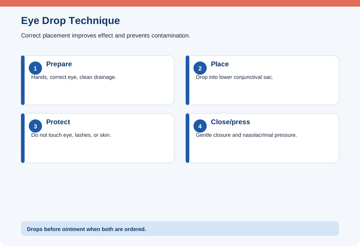
Perform hand hygiene and verify the correct eye. Clean drainage from inner to outer canthus with a separate area of cloth for each stroke. Ask the patient to look up, pull down the lower lid, and place drops in the conjunctival sac—not directly on the cornea. Do not touch the dropper tip to the eye, lashes, skin, or fingers.
After the drop, ask the patient to close the eye gently without squeezing. Apply light pressure to the nasolacrimal duct at the inner canthus for about one minute or as instructed to reduce systemic absorption. When more than one drop is ordered, allow the recommended interval; administer drops before ointment so ointment does not block absorption.
For ointment, apply a thin ribbon from inner to outer conjunctival sac. Temporary blurred vision is expected, so use fall precautions. Never share eye medication between patients.
Communicate with hearing loss
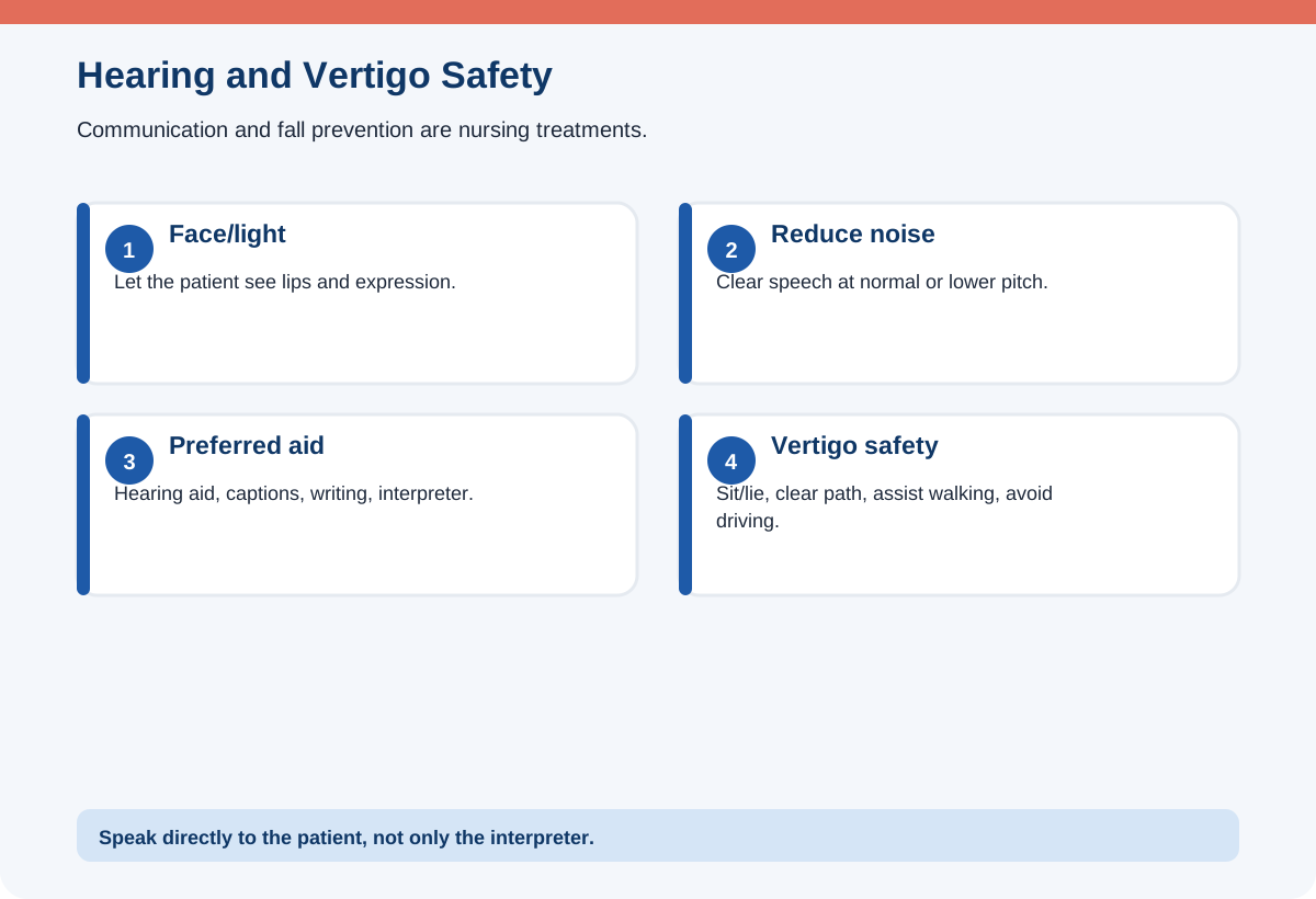
Determine the patient’s preferred method: hearing aids, lip reading, sign language, writing, captioning, or an interpreter. Face the patient in good light, keep hands away from the mouth, reduce background noise, speak clearly at a normal or slightly lower pitch, and use short sentences. Rephrase rather than repeating the same misunderstood words louder.
Check that hearing aids are inserted, powered, clean, and have working batteries. Protect them from water, heat, and loss. Before procedures, store them in a labeled container if removal is necessary, but leave them in whenever communication and safety benefit.
For a person who uses sign language, use a qualified interpreter for informed consent and important education. Speak directly to the patient, not to the interpreter. Do not rely on family for complex medical interpretation when professional services are available.
Understand Ménière disease, otitis, and vestibular disorders
Ménière disease commonly causes episodic vertigo, fluctuating sensorineural hearing loss, tinnitus, and ear fullness. During an attack, protect from falls, reduce motion and stimulation, assist with mobility, and administer vestibular suppressants or antiemetics as ordered. Long-term management may include sodium reduction, avoidance of personal triggers, and prescribed diuretics or other therapy.
Benign paroxysmal positional vertigo causes brief vertigo with position changes and may be treated with canalith-repositioning maneuvers. Vestibular neuritis causes acute prolonged vertigo without hearing loss, while labyrinthitis may include hearing symptoms. Sudden neurologic deficits, severe headache, inability to walk, or new unilateral weakness require stroke evaluation rather than a simple vertigo label.
Otitis externa causes ear-canal pain, often with tragus or pinna tenderness. Otitis media involves the middle ear and may follow upper-respiratory infection. Keep the ear dry as directed and complete prescribed therapy. Mastoid tenderness, swelling behind the ear, facial weakness, or systemic illness requires urgent assessment.
Administer ear medications and protect safety
Warm ear drops in the hands to reduce dizziness, verify the affected ear, position it upward, and straighten the canal. For adults and older children, pull the pinna up and back; for children younger than about three years, pull down and back. Instill along the canal wall without touching the dropper and keep the patient positioned for several minutes.
Do not place drops into an ear with suspected perforation unless the product is specifically approved and ordered. Severe pain, drainage after trauma, sudden hearing loss, or a foreign body requires evaluation. Do not irrigate a button battery, vegetable matter that can swell, or an ear with possible perforation.
Vertigo creates fall risk. Keep the bed low, call light available, pathways clear, and assist with ambulation. Teach the patient to rise slowly, avoid driving during attacks, and sit or lie down at the first warning.
Clinical integration: connecting the topic to a complete patient picture
Sensory disorders can create secondary safety problems. A patient with peripheral-field loss from glaucoma may bump into objects even when central vision is clear. Approach from the better-seeing side, keep frequently used items in a consistent location, improve lighting without glare, and teach scanning of the environment. A patient with reduced hearing may appear confused when the actual problem is that instructions were delivered from behind in a noisy room.
Medication reconciliation is also important. Some eye drops have systemic effects, and some systemic medications can worsen pressure or vestibular symptoms. Ask about asthma, bradycardia, heart block, urinary retention, and current medications before administering or teaching ophthalmic therapy. During ear and eye procedures, confirm laterality repeatedly because wrong-side treatment can cause direct harm.
Discharge teaching should be functional. Instead of only listing drops, ask the patient to demonstrate hand hygiene, bottle identification, conjunctival-sac placement, punctal pressure, spacing, and storage. For hearing aids, verify battery management and safe storage. For vertigo, rehearse how to sit or lie down immediately and how to request assistance. Teach-back reveals barriers that a simple “Do you understand?” will miss.
A simple NCLEX® clinical-judgment workflow
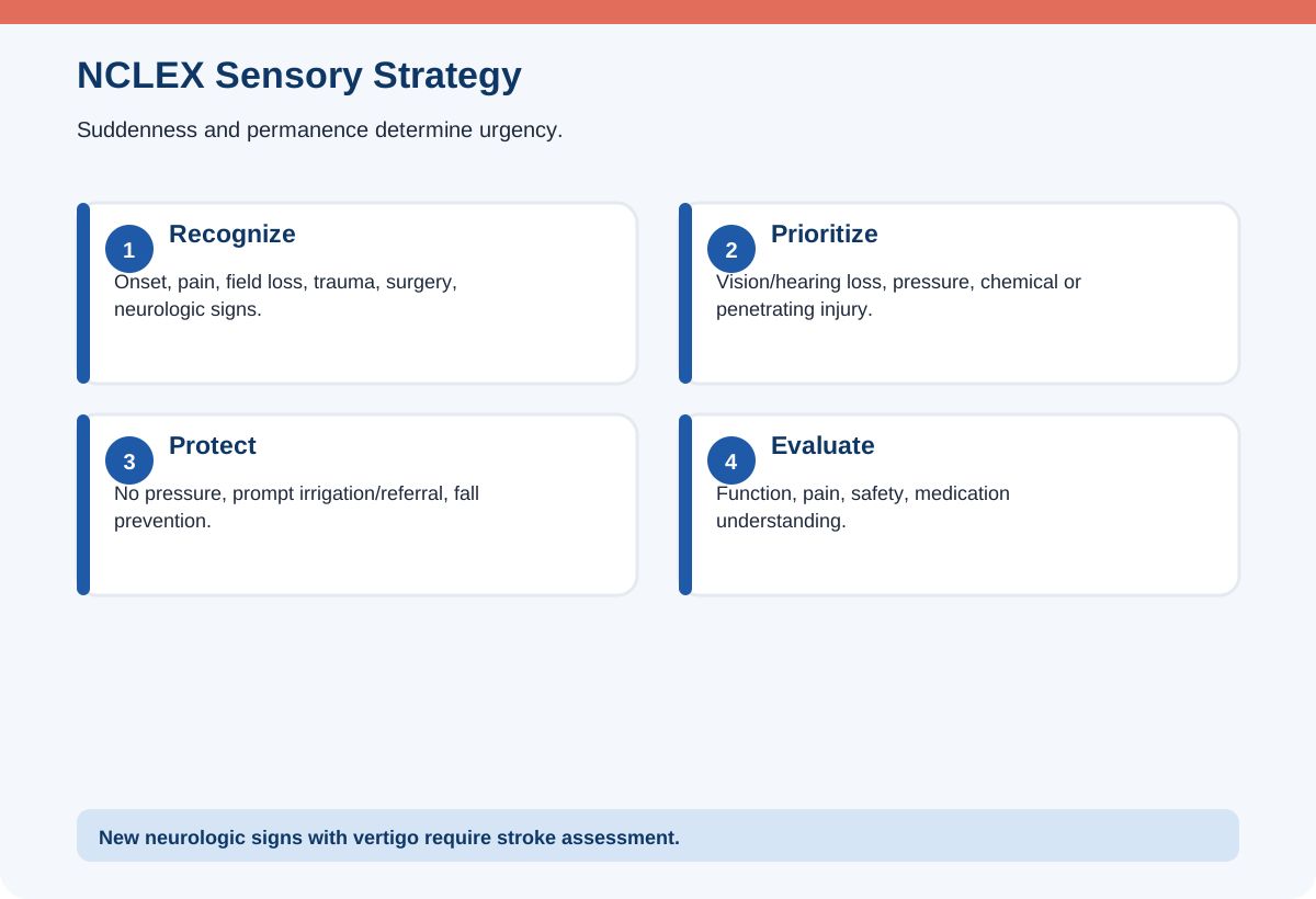
Recognize onset, pain, laterality, trauma, visual-field change, hearing pattern, vertigo, neurologic signs, medication effects, and recent surgery. Analyze whether the disorder is gradual or sudden, sensory or neurologic, and whether pressure, retinal tissue, or airway/fall safety is threatened. Prioritize chemical injury, open globe, angle closure, retinal detachment, sudden hearing loss, and stroke-like vertigo. Generate protective actions and urgent referral. Take action without pressure, delay, or unsafe irrigation. Evaluate vision/hearing, pain, pressure symptoms, mobility, and treatment understanding.
Original practice scenarios with rationales
These are original educational examples written for RN Clarity. They are not copied, recalled, or represented as actual NCLEX® questions.
Scenario 1: Curtain over vision
A patient reports sudden flashes, many new floaters, and a dark curtain over one eye.
Best response: Arrange urgent ophthalmic evaluation for possible retinal detachment.
Rationale: The classic painless pattern can progress to permanent vision loss.
Why the alternatives are weaker: Scheduling a routine eye exam delays care. Irrigating the eye does not treat retinal separation.
Scenario 2: Acute painful red eye
A patient has severe eye pain, halos, headache, nausea, and a red eye.
Best response: Treat as acute angle-closure glaucoma and obtain immediate pressure-lowering care.
Rationale: The optic nerve is at risk from rapidly increased pressure.
Why the alternatives are weaker: Applying an eye patch and waiting is unsafe. Mydriatic drops may worsen the angle.
Scenario 3: Post-cataract pain
One day after cataract surgery, a patient reports severe eye pain and sudden decreased vision.
Best response: Notify the ophthalmic surgeon immediately.
Rationale: Severe pain and vision loss are not expected and may indicate serious postoperative complication.
Why the alternatives are weaker: Routine analgesic and reassessment later can delay treatment. Rubbing the eye is unsafe.
Scenario 4: Gas bubble teaching
A patient with a retinal gas bubble plans to fly home tomorrow.
Best response: Tell the patient not to fly until the ophthalmologist confirms the gas bubble is fully absorbed.
Rationale: Altitude can expand the bubble and dangerously raise pressure.
Why the alternatives are weaker: An eye patch does not prevent expansion. Extra drops do not make air travel safe.
Scenario 5: Ménière attack
A patient with Ménière disease develops sudden spinning, nausea, and unsteady gait.
Best response: Assist the patient to lie safely, reduce stimulation, use fall precautions, and give prescribed symptom medication.
Rationale: Preventing injury and reducing vestibular stimulation are immediate priorities.
Why the alternatives are weaker: Encouraging walking increases fall risk. Rapid head movement worsens vertigo.
Common NCLEX® traps
- All painless vision loss is nonurgent. Retinal detachment and vascular events may be painless emergencies.
- Severe pain is expected after cataract surgery. It requires urgent evaluation.
- Touch dropper to the eye. This contaminates the medication and can injure tissue.
- Skip punctal occlusion. Systemic effects can occur from eye drops.
- Shout at hearing loss. Loudness can distort speech; clear face-to-face communication is better.
- All vertigo is inner-ear disease. Neurologic red flags require stroke evaluation.
- Fly with retinal gas bubble. Altitude expansion can cause dangerous pressure.
- Irrigate every ear foreign body. Batteries, swelling material, and perforation are contraindications.
What to memorize and what to understand
Memorize the red flags for retinal detachment, acute angle closure, chemical exposure, open-globe injury, postoperative cataract complications, and sudden hearing loss. Understand medication administration, punctal occlusion, gas-bubble restrictions, and sensory communication.
Use the “sudden, severe, shadow, splash, surgery” rule: sudden loss, severe pain, a shadow/curtain, chemical splash, or severe symptoms after surgery are urgent.
A seven-day review plan
Day 1: Compare glaucoma, cataract, retinal detachment, and macular degeneration. Day 2: Study chemical and penetrating-eye emergencies. Day 3: Practice eye-drop and ointment technique. Day 4: Review cataract and retinal postoperative teaching. Day 5: Practice communication for hearing loss and hearing-aid care. Day 6: Compare Ménière disease, BPPV, labyrinthitis, and neurologic vertigo. Day 7: Complete mixed sensory scenarios and identify which finding threatens permanent function or immediate safety.
Frequently asked questions
What are classic retinal-detachment symptoms?
Sudden flashes, many new floaters, and a curtain or shadow over part of the visual field.
What makes acute angle-closure glaucoma urgent?
Rapid pressure rise can permanently damage the optic nerve and commonly causes severe pain, halos, nausea, and a red eye.
How can systemic absorption of eye drops be reduced?
Close the eye gently and apply light pressure to the nasolacrimal duct after instillation.
Can a patient fly with a gas bubble in the eye?
No, not until the ophthalmologist confirms it is absorbed, because altitude can expand the gas.
How should a nurse communicate with hearing loss?
Face the patient, reduce noise, speak clearly at a normal/lower pitch, use the preferred aid or interpreter, and verify understanding.
What is the priority during a Ménière attack?
Prevent falls, reduce motion and stimulation, and treat severe vertigo and nausea as ordered.
Final rapid-review checklist
- I recognize retinal detachment and acute angle closure.
- I irrigate chemical eye exposure immediately.
- I never remove an impaled eye object or apply pressure.
- I know severe pain after cataract surgery is abnormal.
- I administer eye drops into the conjunctival sac and use punctal pressure.
- I know gas-bubble altitude restrictions.
- I communicate effectively with hearing impairment.
- I use fall precautions and check stroke red flags with vertigo.
Sources and further reading
- NCSBN, 2026 NCLEX-RN® Test Plan: https://www.nclex.com/test-plans.page
- National Eye Institute, Retinal Detachment: https://www.nei.nih.gov/learn-about-eye-health/eye-conditions-and-diseases/retinal-detachment
- National Eye Institute, Glaucoma: https://www.nei.nih.gov/learn-about-eye-health/eye-conditions-and-diseases/glaucoma
- National Eye Institute, Cataracts: https://www.nei.nih.gov/learn-about-eye-health/eye-conditions-and-diseases/cataracts
- NIDCD, Ménière’s Disease: https://www.nidcd.nih.gov/health/menieres-disease
- NIDCD, Sudden Deafness: https://www.nidcd.nih.gov/health/sudden-deafness
Educational disclaimer
RN Clarity provides educational study support only. This article is not medical advice, does not replace a nursing program, clinical instructor, employer policy, or current provider order, and is not affiliated with or endorsed by NCSBN, Pearson VUE, or the NCLEX-RN® program. In an actual clinical setting, follow current laws, facility policies, approved references, and the directions of the responsible licensed clinician.
Structured data
{
  "@context": "https://schema.org",
  "@graph": [
    {
      "@type": "BlogPosting",
      "headline": "NCLEX-RN® Eye and Ear Disorders: Glaucoma, Cataracts, Retinal Detachment, Hearing Loss, and Ménière Disease",
      "description": "Learn glaucoma, cataracts, retinal detachment, eye emergencies, hearing loss, ear medications, Ménière disease, and nursing safety for NCLEX®.",
      "mainEntityOfPage": {
        "@type": "WebPage",
        "@id": "https://rnclarity.com/blog/nclex-rn-eye-ear-disorders-glaucoma-retinal-detachment"
      },
      "url": "https://rnclarity.com/blog/nclex-rn-eye-ear-disorders-glaucoma-retinal-detachment",
      "datePublished": "ACTUAL_FIRST_LIVE_DATE",
      "dateModified": "ACTUAL_FIRST_LIVE_DATE",
      "author": {
        "@type": "Organization",
        "name": "RN Clarity"
      },
      "publisher": {
        "@type": "Organization",
        "name": "RN Clarity"
      },
      "inLanguage": "en-US",
      "isAccessibleForFree": true,
      "about": [
        "NCLEX-RN® examination",
        "Nursing education",
        "NCLEX® eye and ear disorders"
      ]
    },
    {
      "@type": "BreadcrumbList",
      "itemListElement": [
        {
          "@type": "ListItem",
          "position": 1,
          "name": "Home",
          "item": "https://rnclarity.com/"
        },
        {
          "@type": "ListItem",
          "position": 2,
          "name": "Blog",
          "item": "https://rnclarity.com/blog"
        },
        {
          "@type": "ListItem",
          "position": 3,
          "name": "NCLEX-RN® Eye and Ear Disorders: Glaucoma, Cataracts, Retinal Detachment, Hearing Loss, and Ménière Disease",
          "item": "https://rnclarity.com/blog/nclex-rn-eye-ear-disorders-glaucoma-retinal-detachment"
        }
      ]
    },
    {
      "@type": "FAQPage",
      "mainEntity": [
        {
          "@type": "Question",
          "name": "What are classic retinal-detachment symptoms?",
          "acceptedAnswer": {
            "@type": "Answer",
            "text": "Sudden flashes, many new floaters, and a curtain or shadow over part of the visual field."
          }
        },
        {
          "@type": "Question",
          "name": "What makes acute angle-closure glaucoma urgent?",
          "acceptedAnswer": {
            "@type": "Answer",
            "text": "Rapid pressure rise can permanently damage the optic nerve and commonly causes severe pain, halos, nausea, and a red eye."
          }
        },
        {
          "@type": "Question",
          "name": "How can systemic absorption of eye drops be reduced?",
          "acceptedAnswer": {
            "@type": "Answer",
            "text": "Close the eye gently and apply light pressure to the nasolacrimal duct after instillation."
          }
        },
        {
          "@type": "Question",
          "name": "Can a patient fly with a gas bubble in the eye?",
          "acceptedAnswer": {
            "@type": "Answer",
            "text": "No, not until the ophthalmologist confirms it is absorbed, because altitude can expand the gas."
          }
        },
        {
          "@type": "Question",
          "name": "How should a nurse communicate with hearing loss?",
          "acceptedAnswer": {
            "@type": "Answer",
            "text": "Face the patient, reduce noise, speak clearly at a normal/lower pitch, use the preferred aid or interpreter, and verify understanding."
          }
        },
        {
          "@type": "Question",
          "name": "What is the priority during a Ménière attack?",
          "acceptedAnswer": {
            "@type": "Answer",
            "text": "Prevent falls, reduce motion and stimulation, and treat severe vertigo and nausea as ordered."
          }
        }
      ]
    }
  ]
}