NCLEX-RN® Fetal Heart Rate Monitoring: Baseline, Variability, Accelerations, and Decelerations
Learn fetal heart-rate baseline, variability, accelerations, decelerations, and priority nursing responses.
postNumber: 83 title: "NCLEX-RN® Fetal Heart Rate Monitoring: Baseline, Variability, Accelerations, and Decelerations" h1: "NCLEX-RN® Fetal Heart Rate Monitoring: Baseline, Variability, Accelerations, and Decelerations" slug: "nclex-rn-fetal-heart-rate-monitoring-decelerations" canonicalUrl: "https://rnclarity.com/blog/nclex-rn-fetal-heart-rate-monitoring-decelerations" primaryKeyword: "NCLEX® fetal heart rate monitoring" secondaryKeywords:
- "early late variable decelerations NCLEX®"
- "fetal heart rate variability nursing"
- "category I II III fetal tracing"
- "intrauterine resuscitation nursing"
- "labor fetal monitoring guide" metaTitle: "NCLEX® Fetal Heart Monitoring: Decelerations Guide" metaDescription: "Learn fetal heart baseline, variability, accelerations, early, variable, late, and prolonged decelerations plus safe nursing responses for NCLEX®." excerpt: "A plain-language guide to reading fetal heart patterns, linking them to physiology, and choosing safe first nursing actions." suggestedPublishDate: "2026-08-22" status: "draft_nurse_review_required" schemaTypes: ["BlogPosting", "FAQPage", "BreadcrumbList"] diagramCount: 5 clinicalReview: "Required before publication"
NCLEX-RN® Fetal Heart Rate Monitoring: Baseline, Variability, Accelerations, and Decelerations
Quick answer: Read a fetal tracing in the same order: baseline, variability, accelerations, decelerations, contractions, and the overall clinical picture. Early decelerations usually mirror contractions and suggest head compression; variable decelerations suggest cord compression; late decelerations begin after the contraction starts and suggest reduced uteroplacental oxygen transfer.
Editorial status: This is a complete educational draft. A qualified U.S. registered nurse or nurse educator should clinically review it before publication. Drug doses, facility procedures, and emergency algorithms must be checked against the current source and local policy.
Suggested reading time: 18–22 minutes
Designed for: NCLEX-RN® candidates, including internationally educated nurses and repeat test-takers.
Table of contents
- Use a consistent tracing-reading sequence
- Understand normal baseline and variability
- Interpret accelerations
- Interpret early decelerations
- Interpret variable decelerations
- Interpret late and prolonged decelerations
- Recognize uterine tachysystole and medication effects
- Use Category I, II, and III safely
- A simple NCLEX® clinical-judgment workflow
- Original practice scenarios with rationales
- Common NCLEX® traps
- What to memorize and what to understand
- A seven-day review plan
- Frequently asked questions
- Final rapid-review checklist
Why this topic matters for the NCLEX-RN®
Fetal monitoring questions test pattern recognition and rapid nursing response. The tracing is not interpreted from one dip alone. The nurse considers baseline, variability, recurrent patterns, contraction frequency, labor progress, medications, maternal vital signs, and whether the fetus recovers between events.
Modern intrapartum management emphasizes targeted intrauterine resuscitation rather than automatic rituals. Repositioning, reducing uterine stimulation, correcting maternal hypotension, giving an IV fluid bolus when appropriate, treating tachysystole, and escalating care are common actions. Routine oxygen for every abnormal tracing is not supported when the mother is not hypoxemic; follow current policy and the clinical situation.
Use a consistent tracing-reading sequence
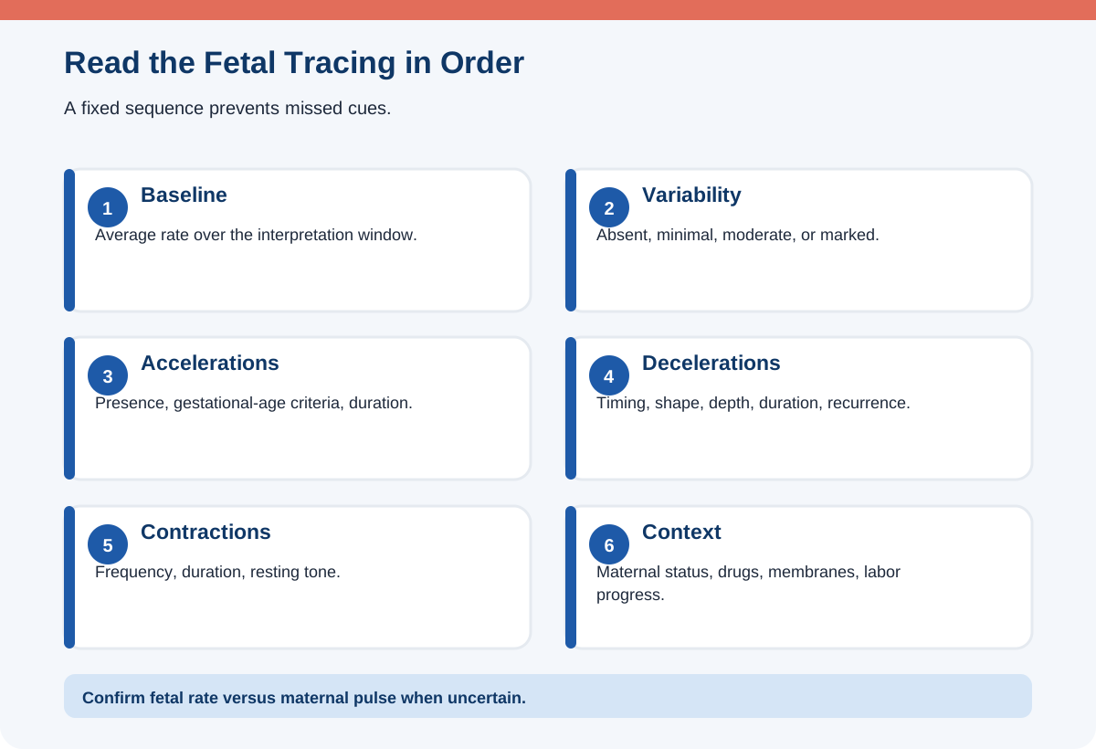
Start with the fetal heart rate baseline over a ten-minute window, excluding major accelerations and decelerations. Then assess variability, accelerations, decelerations, and uterine activity. Finally, connect the pattern to maternal blood pressure, temperature, oxygenation, medications, cervical change, membrane status, and fetal risk factors.
A common normal baseline at term is 110–160 beats/minute. Bradycardia or tachycardia is interpreted by duration and cause. Maternal fever, infection, medications, dehydration, or fetal stress can contribute to tachycardia. Maternal hypotension, cord compression, rapid descent, medications, or fetal compromise can contribute to bradycardia.
Confirm that the monitor is recording the fetal rate rather than the maternal pulse, especially when the rates are similar or the signal changes. Palpate or electronically compare the maternal pulse when there is doubt.
Understand normal baseline and variability
Variability is the beat-to-beat fluctuation around the baseline. Moderate variability, commonly 6–25 beats/minute, is a reassuring sign that the fetal autonomic nervous system is functioning and that significant metabolic acidemia is unlikely at that moment. Minimal variability is detectable but 5 beats/minute or less. Absent variability means no detectable fluctuation. Marked variability exceeds 25 beats/minute.
Minimal variability can occur with fetal sleep, medications, prematurity, or evolving hypoxia. It becomes more concerning when persistent or combined with recurrent late or variable decelerations. Absent variability with recurrent late decelerations, recurrent variable decelerations, bradycardia, or a sinusoidal pattern is highly concerning and requires urgent evaluation.
Do not call a tracing reassuring because the baseline is normal if variability is absent and decelerations are recurrent. Conversely, a brief period of minimal variability may improve with stimulation or time if the fetus is sleeping.
| Feature | Meaning | NCLEX® interpretation |
|---|---|---|
| Baseline 110–160 | Common normal term range | Interpret with variability and decelerations |
| Moderate variability | 6–25 bpm fluctuation | Reassuring at that time |
| Minimal variability | 1–5 bpm fluctuation | Sleep/medication or possible compromise |
| Absent variability | No detectable fluctuation | Concerning with recurrent decelerations/bradycardia |
| Marked variability | >25 bpm fluctuation | May require evaluation in context |
Interpret accelerations
An acceleration is an abrupt increase above baseline. At 32 weeks or more, a common definition is at least 15 beats/minute above baseline lasting at least 15 seconds but less than two minutes. Before 32 weeks, 10 beats/minute for 10 seconds is often used. A prolonged acceleration lasts two minutes or more but less than ten minutes; a change lasting ten minutes becomes a new baseline.
Accelerations generally indicate intact fetal oxygenation and neurologic responsiveness. They may occur with movement, stimulation, or contractions. Their presence is reassuring, but their absence during labor is not automatically abnormal when other features are reassuring.
In antepartum nonstress testing, accelerations are central to a reactive interpretation. The exam may use the gestational-age distinction, so read the stem carefully.
Interpret early decelerations
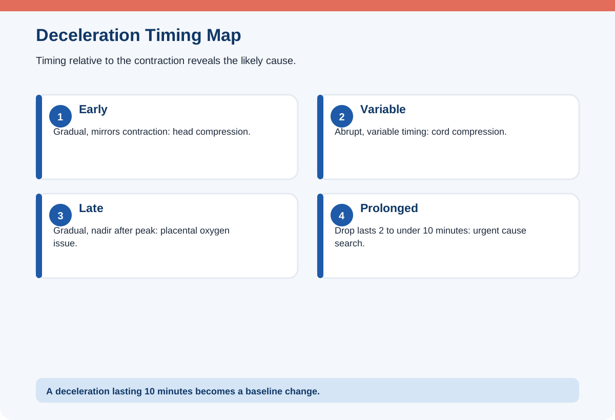
Early decelerations are gradual decreases that mirror the contraction. The nadir occurs near the peak of the contraction, and recovery occurs as the contraction ends. They are commonly caused by fetal head compression and vagal stimulation during labor.
Early decelerations are usually benign when the rest of the tracing is reassuring. Continue observation, assess labor progress, and avoid unnecessary intervention. They often appear as the cervix dilates and the head descends.
The classic memory phrase “early equals head” is useful, but look at timing and shape. A gradual deceleration that starts after the contraction and recovers after it ends is late, not early, even if the fetus is low in the pelvis.
Interpret variable decelerations
Variable decelerations are abrupt decreases that can vary in timing, depth, and shape. They commonly indicate umbilical cord compression. Initial actions include changing maternal position to reduce compression, assessing for cord prolapse when appropriate, reducing uterine stimulation, and notifying or escalating when recurrent or severe.
After rupture of membranes, a sudden prolonged variable or bradycardia requires immediate vaginal assessment for cord prolapse according to the situation. Recurrent variable decelerations may be treated with amnioinfusion in selected patients under an order when low fluid contributes to cord compression.
Evaluate recovery, variability between decelerations, and additional features such as slow return to baseline, overshoots, or increasing depth and duration. Not every variable deceleration indicates severe compromise, but recurrent worsening variables require intervention.
Interpret late and prolonged decelerations
Late decelerations are gradual decreases that begin after the contraction starts; the nadir follows the peak, and recovery occurs after the contraction ends. They suggest reduced uteroplacental oxygen transfer. Causes include maternal hypotension, uterine tachysystole, placental dysfunction, hypertensive disease, diabetes-related placental disease, or abruption.
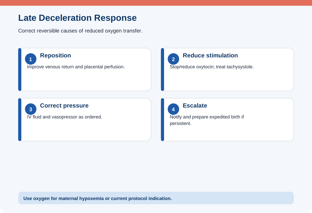
Reposition the patient, stop or reduce oxytocin according to protocol, correct hypotension, provide an IV fluid bolus when appropriate, treat tachysystole, assess maternal oxygenation, and notify the provider. Give supplemental oxygen when the mother is hypoxemic or when directed by the current emergency protocol rather than automatically for every late deceleration.
A prolonged deceleration lasts at least two minutes but less than ten minutes. Search quickly for cord prolapse, hypotension, tachysystole, rapid descent, abruption, uterine rupture, or medication effect. Persistent bradycardia or a prolonged deceleration that does not recover can require expedited birth.
Recognize uterine tachysystole and medication effects
Tachysystole generally means more than five contractions in ten minutes averaged over thirty minutes. It can reduce fetal recovery time and uteroplacental blood flow. Assess contraction frequency, duration, resting tone, and fetal response.
When oxytocin is infusing and tachysystole occurs with an abnormal fetal pattern, stop or reduce the infusion according to protocol, reposition, give fluids when appropriate, and notify. A tocolytic may be ordered if contractions do not resolve. Never increase oxytocin when the fetus is showing recurrent late decelerations or the uterus is overstimulated.
Epidural-related hypotension can produce late decelerations. Positioning, IV fluid, and vasopressor therapy may be ordered. Maternal fever can cause fetal tachycardia; treat the maternal cause rather than focusing only on the monitor.
Use Category I, II, and III safely
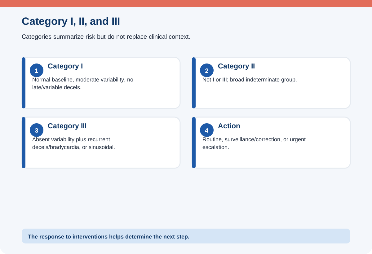
Category I is normal and includes a baseline of 110–160, moderate variability, no late or variable decelerations, with early decelerations and accelerations either present or absent. Continue routine monitoring.
Category III includes absent variability with recurrent late decelerations, recurrent variable decelerations, or bradycardia, or a sinusoidal pattern. It requires prompt evaluation and attempts to correct reversible causes; if unresolved, expedited delivery may be necessary.
Category II includes everything that is not Category I or III. It is broad and requires ongoing surveillance, clinical context, corrective measures, and reassessment. Do not treat “Category II” as a diagnosis or automatically assume immediate cesarean birth. The trend and response to interventions matter.
Clinical integration: connecting the topic to a complete patient picture
A complete fetal-monitoring question usually includes more than a tracing label. Imagine a patient at 39 weeks receiving oxytocin after an epidural. The fetal baseline is initially 140 with moderate variability. The maternal blood pressure falls, contractions become frequent, and recurrent late decelerations appear. The safest answer is not simply “late decelerations equal placental insufficiency.” It is to recognize a reversible combination: maternal hypotension and uterine tachysystole are reducing placental oxygen transfer. Repositioning, reducing oxytocin, correcting blood pressure and intravascular volume as ordered, and reassessing the tracing form one coherent plan.
Now change one cue. If membranes just ruptured and the fetal rate suddenly falls to 70, cord prolapse moves to the top of the differential. The nurse calls for help, performs the indicated vaginal assessment, relieves pressure from the presenting part if a cord is found, positions to reduce compression, and prepares emergency birth. The same low heart rate has a different cause and action because the timeline changed.
Documentation should include the tracing feature, onset, duration, relationship to contractions, maternal assessment, interventions, notifications, and response. Avoid vague notes such as “fetal distress.” Describe the observable pattern. Clear documentation helps the team see whether the fetus recovered and whether the plan must escalate.
A simple NCLEX® clinical-judgment workflow
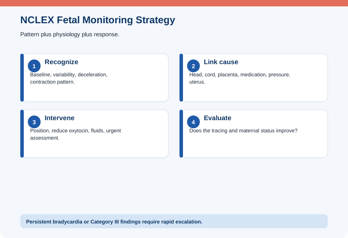
Recognize baseline, variability, acceleration and deceleration timing, contraction pattern, oxytocin use, membrane status, maternal blood pressure and temperature, and labor stage. Analyze the likely physiology and whether the fetus recovers. Prioritize prolonged bradycardia, absent variability with recurrent decelerations, cord prolapse, abruption, rupture, and severe tachysystole. Generate reversible-cause interventions while preparing escalation. Take action and document the time and response. Evaluate whether variability, baseline, decelerations, and maternal status improve.
Original practice scenarios with rationales
These are original educational examples written for RN Clarity. They are not copied, recalled, or represented as actual NCLEX® questions.
Scenario 1: Late decelerations on oxytocin
A patient receiving oxytocin develops recurrent gradual decelerations whose nadirs occur after contraction peaks.
Best response: Reposition, stop or reduce oxytocin per protocol, assess maternal blood pressure and oxygenation, give ordered fluids, and notify.
Rationale: The timing indicates late decelerations and oxytocin may be contributing to excessive uterine activity.
Why the alternatives are weaker: Increasing oxytocin worsens oxygen transfer. Waiting for cervical change delays corrective action.
Scenario 2: Abrupt variables after rupture
Immediately after membrane rupture, the fetal rate drops abruptly and remains low.
Best response: Call for help and assess immediately for cord prolapse while initiating intrauterine resuscitation.
Rationale: Sudden bradycardia after rupture is a classic cord-prolapse emergency clue.
Why the alternatives are weaker: A routine position change without vaginal assessment may miss the prolapsed cord. Leaving to obtain consent delays care.
Scenario 3: Early decelerations
The tracing shows gradual decelerations that mirror contractions with moderate variability.
Best response: Continue observation and assess labor progress.
Rationale: The pattern is consistent with benign head compression.
Why the alternatives are weaker: Emergency cesarean birth is not indicated from this pattern alone. Oxygen is not routinely needed.
Scenario 4: Minimal variability after opioid
After an opioid analgesic, variability becomes minimal for a short period without decelerations.
Best response: Continue close monitoring and reassess while considering medication effect and fetal sleep.
Rationale: Transient minimal variability can follow medication; the entire pattern and duration determine concern.
Why the alternatives are weaker: Declaring fetal acidemia from one short period is unsupported. Ignoring it completely misses the need for reassessment.
Scenario 5: Tachysystole with fetal change
There are six contractions in ten minutes and recurrent variable decelerations during oxytocin infusion.
Best response: Reduce uterine stimulation immediately according to protocol and begin corrective measures.
Rationale: The fetus lacks recovery time and oxytocin is a reversible cause.
Why the alternatives are weaker: Increasing the rate is unsafe. Encouraging pushing may increase stress if birth is not imminent.
Common NCLEX® traps
- Naming the dip without timing. Use onset, nadir, and recovery relative to the contraction.
- Normal baseline equals normal tracing. Variability and decelerations can make the pattern dangerous.
- Routine oxygen for every abnormality. Correct the cause and use oxygen when maternal hypoxemia or protocol indicates.
- Category II equals cesarean. It requires surveillance and response, not one automatic outcome.
- Ignore uterine activity. Tachysystole can be the reversible cause.
- Increase oxytocin despite decelerations. Reduce stimulation when oxygen transfer is compromised.
- Assume no variability always means acidemia. Sleep, medication, prematurity, and duration matter.
- Forget maternal pulse. Confirm the monitor is recording the fetus.
What to memorize and what to understand
Memorize the normal baseline range, variability definitions, acceleration criteria by gestational age, and the timing patterns of early, variable, and late decelerations. Understand the physiology: head compression, cord compression, and reduced placental oxygen transfer. Also understand that the safest action targets a reversible cause and then evaluates the fetal response.
A useful sequence is BLVDC: baseline, variability, decelerations, contractions, and context. Accelerations fit between variability and decelerations when you write a full interpretation.
A seven-day review plan
Day 1: Learn baseline and variability. Day 2: Practice acceleration criteria and antepartum reactive testing. Day 3: Draw early, variable, late, and prolonged decelerations. Day 4: Match each pattern with physiology and first actions. Day 5: Review tachysystole, oxytocin, epidural hypotension, cord prolapse, and abruption. Day 6: Classify sample patterns as Category I, II, or III. Day 7: Complete mixed scenarios and explain the tracing aloud in the same reading order each time.
Frequently asked questions
What is a normal fetal heart baseline?
A common normal term baseline is 110–160 beats/minute, interpreted with variability and decelerations.
What does moderate variability mean?
It generally means 6–25 beats/minute of fluctuation and is reassuring at that time.
What causes variable decelerations?
Umbilical cord compression is the common cause.
What causes late decelerations?
Reduced uteroplacental oxygen transfer, often related to hypotension, tachysystole, or placental dysfunction.
Should oxygen be given for every late deceleration?
Not routinely when the mother is not hypoxemic. Use current policy and target reversible causes such as position, oxytocin, hypotension, and tachysystole.
What makes Category III urgent?
Absent variability with recurrent late or variable decelerations or bradycardia, or a sinusoidal pattern, signals a high-risk pattern requiring prompt correction and possible expedited delivery.
Final rapid-review checklist
- I read baseline, variability, accelerations, decelerations, contractions, and context.
- I distinguish gradual early/late from abrupt variable decelerations.
- I recognize moderate variability as reassuring.
- I stop or reduce oxytocin when tachysystole and fetal compromise occur.
- I assess for cord prolapse after sudden bradycardia following rupture of membranes.
- I do not use routine oxygen as a substitute for correcting causes.
- I understand Category II is broad and trend-based.
- I recognize unresolved Category III patterns as urgent.
Sources and further reading
- NCSBN, 2026 NCLEX-RN® Test Plan: https://www.nclex.com/test-plans.page
- American College of Obstetricians and Gynecologists, Intrapartum Fetal Heart Rate Monitoring: Interpretation and Management: https://www.acog.org/clinical/clinical-guidance/clinical-practice-guideline/articles/2025/10/intrapartum-fetal-heart-rate-monitoring-interpretation-and-management
- ACOG, Fetal Heart Rate Nomenclature, Interpretation, and Management education: https://www.acog.org/education-and-events/emodules/fetal-heart-rate-nomenclature-interpretation-management
- ACOG, Antepartum Fetal Surveillance: https://www.acog.org/clinical/clinical-guidance/practice-bulletin/articles/2021/06/antepartum-fetal-surveillance
Educational disclaimer
RN Clarity provides educational study support only. This article is not medical advice, does not replace a nursing program, clinical instructor, employer policy, or current provider order, and is not affiliated with or endorsed by NCSBN, Pearson VUE, or the NCLEX-RN® program. In an actual clinical setting, follow current laws, facility policies, approved references, and the directions of the responsible licensed clinician.
Structured data
{
  "@context": "https://schema.org",
  "@graph": [
    {
      "@type": "BlogPosting",
      "headline": "NCLEX-RN® Fetal Heart Rate Monitoring: Baseline, Variability, Accelerations, and Decelerations",
      "description": "Learn fetal heart baseline, variability, accelerations, early, variable, late, and prolonged decelerations plus safe nursing responses for NCLEX®.",
      "mainEntityOfPage": {
        "@type": "WebPage",
        "@id": "https://rnclarity.com/blog/nclex-rn-fetal-heart-rate-monitoring-decelerations"
      },
      "url": "https://rnclarity.com/blog/nclex-rn-fetal-heart-rate-monitoring-decelerations",
      "datePublished": "ACTUAL_FIRST_LIVE_DATE",
      "dateModified": "ACTUAL_FIRST_LIVE_DATE",
      "author": {
        "@type": "Organization",
        "name": "RN Clarity"
      },
      "publisher": {
        "@type": "Organization",
        "name": "RN Clarity"
      },
      "inLanguage": "en-US",
      "isAccessibleForFree": true,
      "about": [
        "NCLEX-RN® examination",
        "Nursing education",
        "NCLEX® fetal heart rate monitoring"
      ]
    },
    {
      "@type": "BreadcrumbList",
      "itemListElement": [
        {
          "@type": "ListItem",
          "position": 1,
          "name": "Home",
          "item": "https://rnclarity.com/"
        },
        {
          "@type": "ListItem",
          "position": 2,
          "name": "Blog",
          "item": "https://rnclarity.com/blog"
        },
        {
          "@type": "ListItem",
          "position": 3,
          "name": "NCLEX-RN® Fetal Heart Rate Monitoring: Baseline, Variability, Accelerations, and Decelerations",
          "item": "https://rnclarity.com/blog/nclex-rn-fetal-heart-rate-monitoring-decelerations"
        }
      ]
    },
    {
      "@type": "FAQPage",
      "mainEntity": [
        {
          "@type": "Question",
          "name": "What is a normal fetal heart baseline?",
          "acceptedAnswer": {
            "@type": "Answer",
            "text": "A common normal term baseline is 110–160 beats/minute, interpreted with variability and decelerations."
          }
        },
        {
          "@type": "Question",
          "name": "What does moderate variability mean?",
          "acceptedAnswer": {
            "@type": "Answer",
            "text": "It generally means 6–25 beats/minute of fluctuation and is reassuring at that time."
          }
        },
        {
          "@type": "Question",
          "name": "What causes variable decelerations?",
          "acceptedAnswer": {
            "@type": "Answer",
            "text": "Umbilical cord compression is the common cause."
          }
        },
        {
          "@type": "Question",
          "name": "What causes late decelerations?",
          "acceptedAnswer": {
            "@type": "Answer",
            "text": "Reduced uteroplacental oxygen transfer, often related to hypotension, tachysystole, or placental dysfunction."
          }
        },
        {
          "@type": "Question",
          "name": "Should oxygen be given for every late deceleration?",
          "acceptedAnswer": {
            "@type": "Answer",
            "text": "Not routinely when the mother is not hypoxemic. Use current policy and target reversible causes such as position, oxytocin, hypotension, and tachysystole."
          }
        },
        {
          "@type": "Question",
          "name": "What makes Category III urgent?",
          "acceptedAnswer": {
            "@type": "Answer",
            "text": "Absent variability with recurrent late or variable decelerations or bradycardia, or a sinusoidal pattern, signals a high-risk pattern requiring prompt correction and possible expedited delivery."
          }
        }
      ]
    }
  ]
}