NCLEX-RN® IV Therapy and Central Lines: Fluids, Complications, PICCs, Ports, and Safety
Study IV therapy, central-line safety, PICCs, ports, fluid administration, and complications for NCLEX-RN®.
postNumber: 80 title: "NCLEX-RN® IV Therapy and Central Lines: Fluids, Complications, PICCs, Ports, and Safety" h1: "NCLEX-RN® IV Therapy and Central Lines: Fluids, Complications, PICCs, Ports, and Safety" slug: "nclex-rn-iv-therapy-central-lines-picc-port-complications" canonicalUrl: "https://rnclarity.com/blog/nclex-rn-iv-therapy-central-lines-picc-port-complications" primaryKeyword: "NCLEX® IV therapy and central lines" secondaryKeywords:
- "IV infiltration versus extravasation"
- "PICC line nursing care"
- "central line complications NCLEX®"
- "phlebitis nursing intervention"
- "CLABSI prevention nursing" metaTitle: "NCLEX® IV Therapy and Central Lines: Safety Guide" metaDescription: "Learn IV fluid principles, infiltration, extravasation, phlebitis, central-line complications, PICCs, ports, air embolism, and CLABSI prevention." excerpt: "A practical NCLEX® guide to peripheral and central IV access, fluid selection, site complications, infection prevention, and emergency actions." suggestedPublishDate: "2026-08-13" status: "draft_nurse_review_required" schemaTypes: ["BlogPosting", "FAQPage", "BreadcrumbList"] diagramCount: 5 clinicalReview: "Required before publication"
NCLEX-RN® IV Therapy and Central Lines: Fluids, Complications, PICCs, Ports, and Safety
Quick answer: For any IV problem, stop the infusion first when continuing could cause harm, assess the site and patient, identify whether the problem is infiltration, extravasation, phlebitis, infection, occlusion, overload, or a central-line emergency, then follow the medication- and device-specific protocol.
Editorial status: This is a complete educational draft. A qualified U.S. registered nurse or nurse educator should clinically review it before publication. Drug doses, facility procedures, and emergency algorithms must be checked against the current source and local policy.
Suggested reading time: 18–22 minutes
Designed for: NCLEX-RN® candidates, including internationally educated nurses and repeat test-takers.
Table of contents
- Understand peripheral and central access
- Choose IV fluids by purpose
- Recognize infiltration and extravasation
- Recognize phlebitis, infection, and occlusion
- Care for PICCs, central venous catheters, and ports
- Prevent central-line bloodstream infection
- Respond to air embolism, thrombosis, and catheter damage
- Monitor systemic fluid and electrolyte complications
- A simple NCLEX® clinical-judgment workflow
- Original practice scenarios with rationales
- Common NCLEX® traps
- What to memorize and what to understand
- A seven-day review plan
- Frequently asked questions
- Final rapid-review checklist
Why this topic matters for the NCLEX-RN®
IV questions test local assessment, systemic assessment, medication safety, and device management at the same time. A cool swollen site suggests infiltration; burning, blistering, or tissue injury with a vesicant suggests extravasation; redness and a palpable cord suggest phlebitis. A central line adds risks such as bloodstream infection, air embolism, thrombosis, catheter migration, and pneumothorax.
The NCLEX-RN® expects the nurse to recognize the pattern and prevent further injury. “Flush harder” is rarely a safe answer when resistance or swelling is present. Removing a catheter immediately may also be wrong during vesicant extravasation because aspiration of the drug or administration of an antidote may require the catheter to remain temporarily. The safe action depends on the complication.
Understand peripheral and central access
A peripheral IV terminates in a peripheral vein and is used for many short-term fluids and medications. Assess the site frequently for pain, redness, swelling, leakage, coolness, warmth, streaking, and patency. A central venous catheter terminates in a large central vein and can support long-term therapy, vesicants, parenteral nutrition, hemodynamic monitoring, or medications that are too irritating for peripheral veins.
A PICC is inserted through a peripheral vein but its tip is central. A tunneled catheter travels under the skin before entering the vein. An implanted port sits under the skin and is accessed with a noncoring needle. Device type affects dressing, access, flushing, clamping, and removal procedures.
Never use a newly placed central line until tip location has been confirmed by the approved method when confirmation is required. Trace every line from the patient to its source before connecting or administering. Label tubing and lumens clearly to prevent wrong-route errors.
Choose IV fluids by purpose
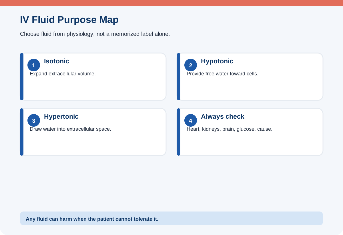
Isotonic crystalloids expand extracellular volume and are commonly used for hypovolemia and initial resuscitation. Examples include 0.9% sodium chloride and balanced isotonic solutions. Large volumes can contribute to overload, edema, and acid-base or electrolyte effects, so reassessment is essential.
Hypotonic fluids provide proportionally more free water and can move water into cells. They may be used in selected hyperosmolar states but can worsen cerebral edema or severe intravascular depletion. Hypertonic fluids draw water into the vascular space and require close monitoring because rapid shifts can cause overload or neurologic injury. Product classification can change after metabolism; for example, a dextrose solution may be isotonic in the bag but physiologically provide free water after glucose is metabolized.
Do not choose a fluid only from the sodium number. Assess blood pressure, perfusion, lung sounds, urine output, neurologic status, glucose, renal function, and the cause of the imbalance. In shock, restoring circulation takes priority; in heart or renal failure, fluid tolerance may be limited.
| Fluid concept | Movement/purpose | Main nursing concern |
|---|---|---|
| Isotonic | Expands extracellular/intravascular volume | Overload and electrolyte/acid-base effects |
| Hypotonic | Provides free water; water moves toward cells | Cerebral edema and worsening intravascular depletion |
| Hypertonic | Pulls water into extracellular space | Rapid shifts, pulmonary edema, neurologic risk |
| Blood products/colloids | Specific replacement or oncotic effect | Reaction, compatibility, and volume tolerance |
Recognize infiltration and extravasation
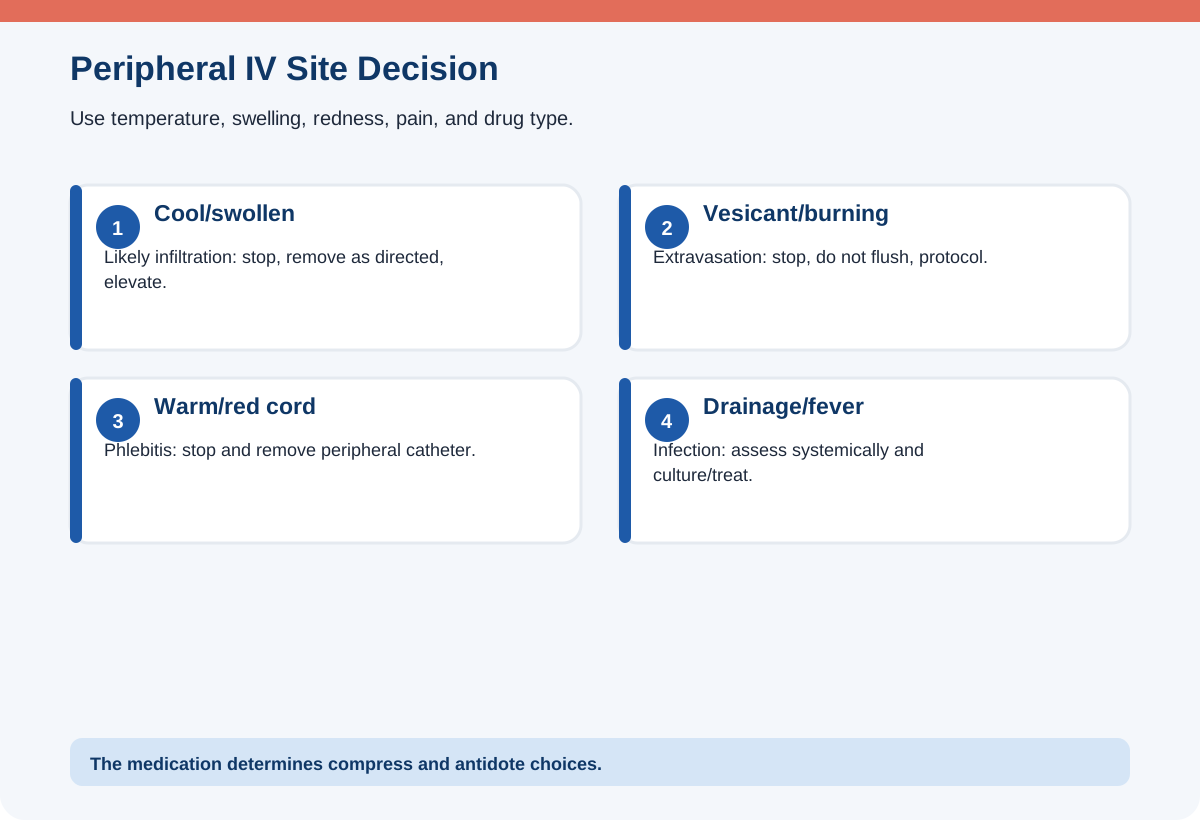
Infiltration occurs when a nonvesicant solution leaks into surrounding tissue. Findings include swelling, coolness, pallor, discomfort, slowed infusion, leakage, and loss of blood return. Stop the infusion, remove the catheter unless a protocol indicates otherwise, elevate the limb, and apply the appropriate compress based on the fluid and policy.
Extravasation is leakage of a vesicant that can cause blistering, necrosis, nerve injury, or loss of function. Stop the infusion immediately. Do not routinely flush, because flushing may spread the drug. Leave the catheter in place initially when the protocol calls for aspiration of residual drug or administration of an antidote. Disconnect the tubing, aspirate as directed, notify the provider and pharmacy, apply the agent-specific warm or cold compress, elevate when appropriate, document, photograph or measure per policy, and arrange follow-up.
The medication determines treatment. Some vesicants require warmth to promote dispersion; others require cold to limit spread. The exam may provide the drug or protocol cue. When it does not, the safest response is to stop and follow the extravasation procedure rather than improvising.
Recognize phlebitis, infection, and occlusion
Phlebitis is vein inflammation. Findings include pain, warmth, redness, swelling, streaking, and sometimes a palpable venous cord. Stop the infusion and remove the peripheral catheter, then restart at a different site if therapy must continue. Apply comfort measures and document the severity according to policy. Purulent drainage, fever, or spreading redness suggests infection and requires further evaluation.
An occlusion may result from a kink, closed clamp, malposition, precipitate, thrombus, or catheter tip against the vessel wall. Check simple mechanical causes first. Never force a flush against resistance. For central lines, use the ordered declotting protocol when a thrombotic occlusion is suspected. Do not use a sharp object to clear tubing.
Bloodstream infection can present with fever, chills, hypotension, confusion, or drainage at the site. Obtain cultures as ordered, begin antimicrobial treatment promptly, and follow the decision about catheter removal. A patient who becomes acutely febrile during central-line infusion needs systemic assessment, not only a dressing change.
Care for PICCs, central venous catheters, and ports
Use strict aseptic technique for access and dressing changes. Scrub the connector for the required time and allow it to dry. Keep dressings clean, dry, intact, and dated according to policy. Replace loose, wet, visibly soiled, or contaminated dressings promptly using the approved antiseptic and barrier precautions.
Confirm blood-return and flushing expectations for the specific device and therapy. Some implanted ports require a noncoring needle and scheduled flushing when not in use. Clamp sequence and positive-pressure technique depend on the connector and device. Follow manufacturer and facility instructions rather than memorizing one universal clamp order.
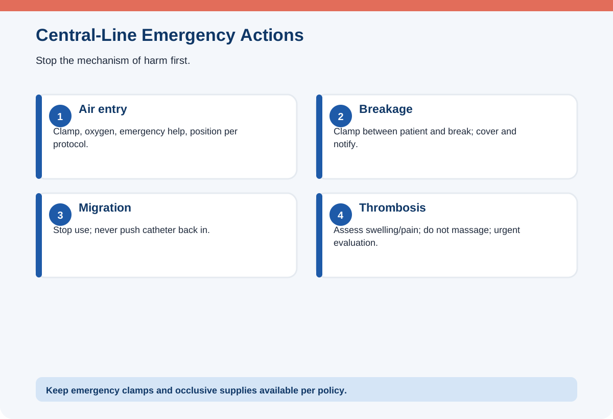
Measure and document external catheter length or other migration markers when required. A change can indicate migration. Do not use scissors near a central catheter. If damage occurs, clamp between the patient and the break with an approved device when possible, cover, and notify urgently. Central-line removal requires a specific position and breath-control technique to reduce air entry.
Prevent central-line bloodstream infection
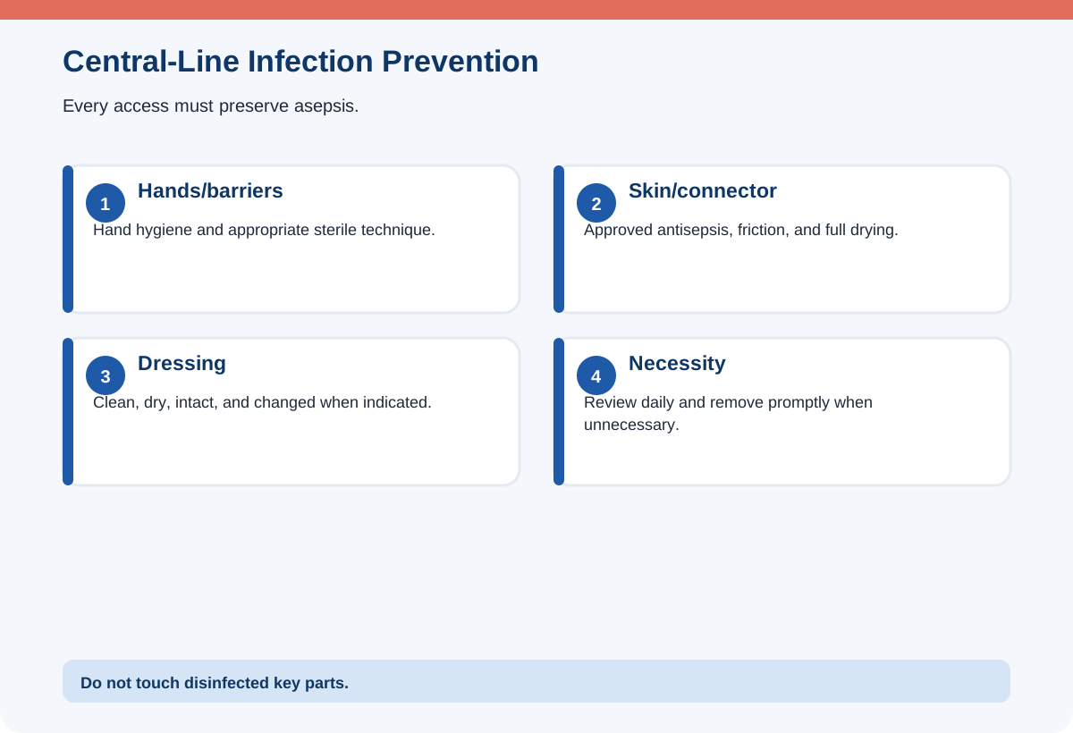
The strongest prevention measures are hand hygiene, aseptic insertion and maintenance, appropriate skin antisepsis, maximal sterile barriers during insertion, daily review of line necessity, and prompt removal when no longer essential. Every access is an opportunity for contamination, so combine medication administration and blood draws when clinically appropriate without compromising therapy.
Disinfect needleless connectors before every access using the required friction and drying time. Do not touch disinfected key parts. Use sterile caps, dressings, and equipment as policy requires. Avoid routine replacement solely by a fixed schedule when evidence-based policy instead uses clinical indication; follow the current institutional standard.
Teach the patient to protect the dressing, avoid submerging the site, report fever, pain, drainage, or loose dressing, and prevent pulling. Infection prevention is not a one-time insertion task; maintenance behavior determines ongoing risk.
Respond to air embolism, thrombosis, and catheter damage
Air can enter a central line through an open hub, disconnected tubing, damaged catheter, or removal process. Sudden dyspnea, chest pain, cough, confusion, hypotension, tachyarrhythmia, or a “mill-wheel” murmur may occur. Clamp the line, stop air entry, call for emergency help, administer oxygen, and position the patient according to current protocol—often left lateral with head down when not contraindicated—to trap air away from the pulmonary outflow while definitive care is arranged.
Central venous thrombosis may cause swelling, pain, venous distention, discoloration, or heaviness in the arm, neck, face, or chest on the catheter side. Stop nonessential infusion and notify for imaging and treatment; do not massage the area.
If a catheter is accidentally removed, apply firm pressure, place an occlusive dressing as directed, assess for air embolism and bleeding, measure the catheter if available to determine whether it appears intact, and notify. Never reinsert a partially dislodged catheter.
Monitor systemic fluid and electrolyte complications
Even a perfect IV site can deliver an unsafe amount or composition. Fluid overload causes dyspnea, crackles, edema, rapid weight gain, rising blood pressure, jugular venous distention, and reduced oxygenation. Stop or slow the infusion as clinically appropriate and obtain urgent evaluation. In contrast, inadequate replacement may leave tachycardia, hypotension, delayed capillary refill, low urine output, and altered mentation.
Monitor glucose when dextrose or parenteral nutrition is used. Abrupt interruption of high-dextrose nutrition can cause hypoglycemia. Central parenteral nutrition also carries infection and electrolyte-shift risks, including refeeding syndrome in severely malnourished patients.
Track intake, output, daily weight, electrolytes, renal function, acid-base status, and cardiopulmonary findings. IV therapy is a treatment that requires ongoing evaluation, not a background task.
A simple NCLEX® clinical-judgment workflow
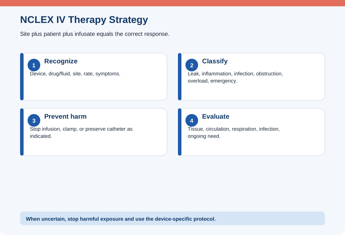
Recognize the device, fluid or medication, site findings, systemic symptoms, recent manipulation, and infusion rate. Analyze whether the problem is tissue leakage, vein inflammation, infection, obstruction, fluid intolerance, thrombosis, or air entry. Prioritize airway compromise, shock, vesicant tissue injury, sepsis, and catheter damage. Generate actions that stop further harm while preserving options such as aspiration through the catheter in extravasation. Take action using the device-specific protocol. Evaluate the limb, tissue, circulation, respiratory status, infection markers, and continued need for the line.
Original practice scenarios with rationales
These are original educational examples written for RN Clarity. They are not copied, recalled, or represented as actual NCLEX® questions.
Scenario 1: Vesicant pain and swelling
During a vesicant infusion, the patient reports burning; the site is swollen and has no blood return.
Best response: Stop the infusion, do not flush, leave the catheter temporarily for aspiration/antidote as directed, and activate the extravasation protocol.
Rationale: Flushing can spread a vesicant and worsen injury.
Why the alternatives are weaker: Increasing the rate is dangerous. Immediate removal without checking the protocol may eliminate the route needed for aspiration.
Scenario 2: Peripheral phlebitis
A peripheral IV site is warm, red, tender, and streaked along the vein.
Best response: Stop the infusion, remove the catheter, assess, and restart elsewhere if needed.
Rationale: The pattern indicates phlebitis and continued infusion worsens inflammation.
Why the alternatives are weaker: A routine flush does not correct inflammation. Covering the site without removal delays care.
Scenario 3: Central-line air entry
After a central-line connection separates, a patient suddenly becomes dyspneic and hypotensive.
Best response: Clamp the line, prevent further air entry, call emergency help, give oxygen, and position per air-embolism protocol.
Rationale: The abrupt event after disconnection is consistent with venous air embolism.
Why the alternatives are weaker: Flushing the catheter can introduce more air. Walking the patient is unsafe.
Scenario 4: Resistance to flushing
A PICC line does not flush easily.
Best response: Stop, check clamps and tubing position, assess the line, and follow the occlusion protocol without force.
Rationale: Force can rupture the catheter or dislodge a thrombus.
Why the alternatives are weaker: Using a smaller syringe to create higher pressure may damage the device. A needle must never be inserted to clear it.
Scenario 5: Fluid overload
A patient with renal impairment receiving isotonic fluid develops crackles, increasing oxygen need, edema, and rising blood pressure.
Best response: Stop or reduce the infusion as appropriate, elevate the head, oxygenate, and notify urgently for overload management.
Rationale: The systemic findings indicate the patient cannot tolerate the infused volume.
Why the alternatives are weaker: A patent IV site does not make the therapy safe. Giving an additional bolus worsens overload.
Common NCLEX® traps
- Flush an infiltrated or extravasated line. Flushing can force more solution into tissue.
- Remove every extravasation catheter immediately. It may be needed briefly for aspiration or antidote administration.
- Force an occluded line. This can rupture the catheter or dislodge material.
- Treat a central line like a peripheral IV. Central access has unique infection, air, migration, and thrombosis risks.
- Choose fluid by sodium alone. Use perfusion, neurologic, renal, cardiac, and cause data.
- Ignore drying time. Antiseptic friction and complete drying are key parts of disinfection.
- Reinsert a dislodged catheter. Never push a migrated catheter back into the body.
- Focus only on the site. Systemic overload or sepsis may occur with a normal-looking site.
What to memorize and what to understand
Memorize the visual and tactile differences among infiltration, extravasation, phlebitis, and infection; the rule not to force a flush; and the emergency sequence for air embolism. Understand why a vesicant catheter may be left briefly, why central-line maintenance prevents infection, and how fluid tonicity changes water movement.
Device-specific details vary, so an exam-safe answer often says to follow the approved protocol after taking the universal first action that prevents further harm.
A seven-day review plan
Day 1: Compare peripheral IVs, PICCs, tunneled catheters, and ports. Day 2: Review isotonic, hypotonic, and hypertonic fluids in clinical scenarios. Day 3: Build a comparison chart for infiltration, extravasation, phlebitis, infection, and occlusion. Day 4: Study connector disinfection, dressing integrity, line necessity, and CLABSI prevention. Day 5: Review air embolism, thrombosis, migration, breakage, and accidental removal. Day 6: Practice five fluid-overload and electrolyte-shift scenarios. Day 7: Complete a mixed set and state the first action, what must not be done, and the reassessment target for each problem.
Frequently asked questions
What is the first action for IV extravasation?
Stop the infusion. Do not flush. Follow the agent-specific protocol, which may require leaving the catheter temporarily for aspiration or antidote.
How is infiltration different from extravasation?
Both are leakage into tissue. Extravasation involves a vesicant capable of serious tissue injury; infiltration involves a nonvesicant solution.
Should resistance in a central line be overcome with force?
No. Check mechanical causes and use the approved occlusion protocol. Never force a flush.
What prevents CLABSI?
Hand hygiene, aseptic insertion and maintenance, connector disinfection with drying, intact dressings, and removing unnecessary lines are central measures.
What does central-line thrombosis look like?
Swelling, pain, heaviness, venous distention, or discoloration of the limb, neck, face, or chest on the catheter side may occur.
Can a partially dislodged central line be pushed back in?
No. Secure it, stop use, and notify for evaluation.
Final rapid-review checklist
- I distinguish infiltration, extravasation, phlebitis, infection, and occlusion.
- I stop a vesicant infusion and do not flush.
- I never force a resistant central line.
- I understand PICC, port, tunneled, and nontunneled access differences.
- I use hand hygiene, aseptic technique, connector scrubbing, and dry time.
- I recognize air embolism, thrombosis, migration, and breakage.
- I monitor the patient for overload and electrolyte effects, not only the site.
- I trace every line before connecting or administering.
Sources and further reading
- NCSBN, 2026 NCLEX-RN® Test Plan: https://www.nclex.com/test-plans.page
- CDC, strategies for preventing central line-associated bloodstream infections: https://www.cdc.gov/infection-control/hcp/intravascular-catheter-related-infection/
- CDC, summary of recommendations for intravascular catheter infection prevention: https://www.cdc.gov/infection-control/hcp/intravascular-catheter-related-infection/summary-recommendations.html
- Infusion Nurses Society, infusion therapy standards information: https://www.ins1.org/
Educational disclaimer
RN Clarity provides educational study support only. This article is not medical advice, does not replace a nursing program, clinical instructor, employer policy, or current provider order, and is not affiliated with or endorsed by NCSBN, Pearson VUE, or the NCLEX-RN® program. In an actual clinical setting, follow current laws, facility policies, approved references, and the directions of the responsible licensed clinician.
Structured data
{
  "@context": "https://schema.org",
  "@graph": [
    {
      "@type": "BlogPosting",
      "headline": "NCLEX-RN® IV Therapy and Central Lines: Fluids, Complications, PICCs, Ports, and Safety",
      "description": "Learn IV fluid principles, infiltration, extravasation, phlebitis, central-line complications, PICCs, ports, air embolism, and CLABSI prevention.",
      "mainEntityOfPage": {
        "@type": "WebPage",
        "@id": "https://rnclarity.com/blog/nclex-rn-iv-therapy-central-lines-picc-port-complications"
      },
      "url": "https://rnclarity.com/blog/nclex-rn-iv-therapy-central-lines-picc-port-complications",
      "datePublished": "ACTUAL_FIRST_LIVE_DATE",
      "dateModified": "ACTUAL_FIRST_LIVE_DATE",
      "author": {
        "@type": "Organization",
        "name": "RN Clarity"
      },
      "publisher": {
        "@type": "Organization",
        "name": "RN Clarity"
      },
      "inLanguage": "en-US",
      "isAccessibleForFree": true,
      "about": [
        "NCLEX-RN® examination",
        "Nursing education",
        "NCLEX® IV therapy and central lines"
      ]
    },
    {
      "@type": "BreadcrumbList",
      "itemListElement": [
        {
          "@type": "ListItem",
          "position": 1,
          "name": "Home",
          "item": "https://rnclarity.com/"
        },
        {
          "@type": "ListItem",
          "position": 2,
          "name": "Blog",
          "item": "https://rnclarity.com/blog"
        },
        {
          "@type": "ListItem",
          "position": 3,
          "name": "NCLEX-RN® IV Therapy and Central Lines: Fluids, Complications, PICCs, Ports, and Safety",
          "item": "https://rnclarity.com/blog/nclex-rn-iv-therapy-central-lines-picc-port-complications"
        }
      ]
    },
    {
      "@type": "FAQPage",
      "mainEntity": [
        {
          "@type": "Question",
          "name": "What is the first action for IV extravasation?",
          "acceptedAnswer": {
            "@type": "Answer",
            "text": "Stop the infusion. Do not flush. Follow the agent-specific protocol, which may require leaving the catheter temporarily for aspiration or antidote."
          }
        },
        {
          "@type": "Question",
          "name": "How is infiltration different from extravasation?",
          "acceptedAnswer": {
            "@type": "Answer",
            "text": "Both are leakage into tissue. Extravasation involves a vesicant capable of serious tissue injury; infiltration involves a nonvesicant solution."
          }
        },
        {
          "@type": "Question",
          "name": "Should resistance in a central line be overcome with force?",
          "acceptedAnswer": {
            "@type": "Answer",
            "text": "No. Check mechanical causes and use the approved occlusion protocol. Never force a flush."
          }
        },
        {
          "@type": "Question",
          "name": "What prevents CLABSI?",
          "acceptedAnswer": {
            "@type": "Answer",
            "text": "Hand hygiene, aseptic insertion and maintenance, connector disinfection with drying, intact dressings, and removing unnecessary lines are central measures."
          }
        },
        {
          "@type": "Question",
          "name": "What does central-line thrombosis look like?",
          "acceptedAnswer": {
            "@type": "Answer",
            "text": "Swelling, pain, heaviness, venous distention, or discoloration of the limb, neck, face, or chest on the catheter side may occur."
          }
        },
        {
          "@type": "Question",
          "name": "Can a partially dislodged central line be pushed back in?",
          "acceptedAnswer": {
            "@type": "Answer",
            "text": "No. Secure it, stop use, and notify for evaluation."
          }
        }
      ]
    }
  ]
}